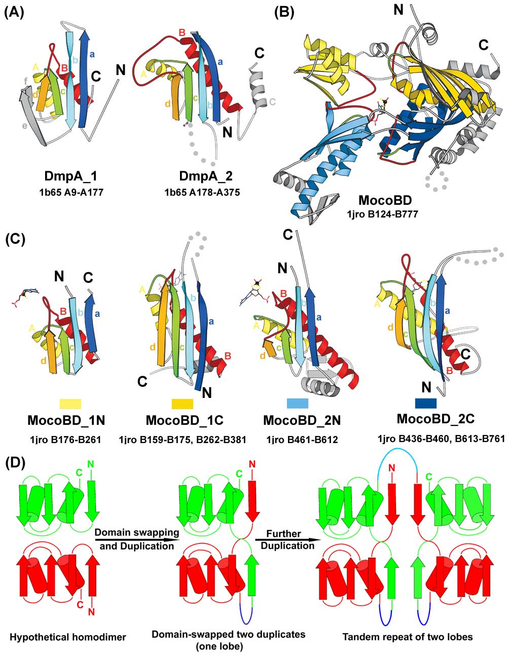
Structural comparison of DmpA and MocoBD domains. (A) The two structural domains of DmpA. (B) The structure of MocoBD. (C) The four domains of MocoBD. In A and C, spatially equivalent secondary structure elements are colored and labeled correspondingly. In B, the four structural domains are shown in different colors: 1N, light yellow; 1C, dark yellow; 2N, light blue; 2C, dark blue. The two lobes are colored in yellow and blue, respectively. The molybdenum cofactor in B and C is shown in bonds representation with the molybdenum atom highlighted as a black ball. In C, the rectangular bar below each structure shows the color in which that domain is represented in B. For all structures, the crossing loops are highlighted in green and red; secondary structure elements that do not belong to the consensus fold of DmpA and MocoBD domains are shown in gray; dotted lines represent disordered regions or long insertions; N and C termini are labeled and the PDB code, the chain ID, and the starting and ending residue numbers are shown. Diagrams are drawn with MOLSCRIPT. (D) A proposed model for MocoBD to evolve from a single-domain ancestor. The single-domain ancestor may form a homodimer (or a homotetramer). Duplication, fusion, and domain swapping created one lobe in which the two duplicates exchange their first strand. The linker region between the two domains is colored in blue. The lower, mainly red domain corresponding to 1N/2N seems to be circularly permuted and inserted into the upper, mainly green domain corresponding to 1C/2C. Further duplication of this lobe resulted in the four domains in MocoBD. The linker between the two lobes is colored in cyan.