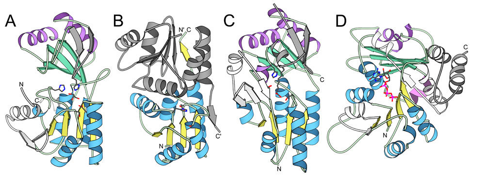
Structural similarities of EDD domain Families. Structural similarities of EDD domain-containing families and tubulin GTPase domain. Structural models produced with MOLSCRIPT reveal similarities between (A) Dak representative structure (1un9) with N-terminal EDD domain, (B) EIIA-man structure (1pdo) of EDD domain swapped dimer, and (C) DegV representative structure (1pzx) with N-terminal EDD domain in comparison to (D) tubulin GTPase domain representative structure (1tub). The α-helices and β-strands of the EDD domain homologs and the N-terminal tubulin domain are colored blue and yellow, respectively. The α-helices and β-strands of the equivalent C-terminal domains are colored purple and green, respectively. Elements inserted into the EDD domain core fold are colored white while elements inserted into the C-terminal domain (or elements corresponding to the EIIA-man swapped dimer that replace the C-terminal domain) are colored gray. Bonds representations of bound ligands (Dihydroxyacetone in Dak and Palmitate in DegV) and family-conserved residues marking the active sites are colored according to atom type (gray for carbon, red for oxygen, blue for nitrogen, and pink for phosphate). The N-terminus and the C-terminus of each structure is labeled.