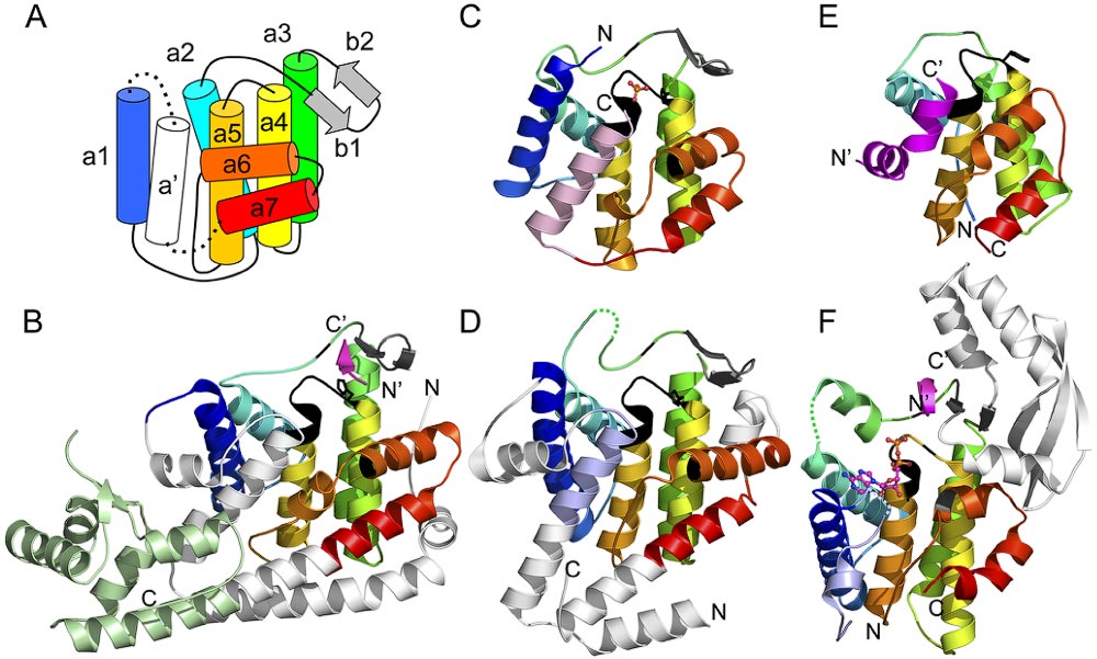
Structural similarities of fido domain-containing families. Structural models of fic homologs define a core fic domain secondary structure topology labeled from N-terminus to C-terminus (A). Diverse fic domain-containing structures are illustrated from a Shewanella oneidensis [2qc0] (B), Helicobacter pylori [2f6s] (C), and Bacteroides thetaiotaomicron [3cuc] (D). The common core a-helices are colored in rainbow from N-terminus (blue) to C-terminus (red). A permuted helix is colored pink (contributed from the C-terminus) or slate (contributed from the N-terminus). Extended elements that decorate the core are colored white, a helix-turn-helix domain is colored light green, and a b-hairpin that binds peptide ligand (magenta) is colored gray. Bound ligands are represented as ball-and-stick and conserved sequence motifs marking the active sites are black. The N-terminus and the C-terminus of each structure is labeled. Similar structures retain most or all of the fic domain core: a doc structure [3dd7] bound to phd antitoxin (magenta) (E) and an AvrB structure bound to Rin4 peptide (magenta) [2nud] also binds ADP (ball-and-stick) [2nun, superimposed] (F).