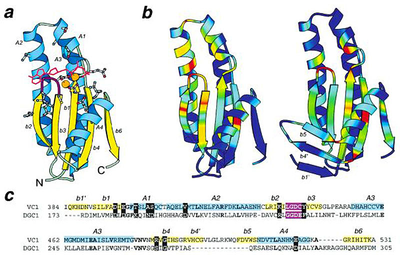
Structural diagrams of GGDEF/cyclase domain: (a) Mammalian adenylyl cyclase catalytic domain (PDB: 1cjv, chain A, residues 384-531) showing the secondary structure elements present in both cyclase catalytic domain and GGDEF domain. Secondary structure elements are labeled by italicized letters and numbers. α-helices and β-strands are labeled with letters A and b, respectively, followed by the corresponding number. GGDEF loop is shown in magenta. Conserved residues in GGDEF domain are shown in ball-and-stick representations of their counterparts in the cyclase catalytic domain. ATP is displayed in red lines; two metal ions are shown as orange balls. Drawn with the program BOBSCRIPT. (b) Ribbon diagrams showing sequence conservation in multiple alignments of GGDEF domain family (left) and cyclase catalytic domain (right). Red and blue correspond to highest and the lowest conservation, respectively. (c) Sequence alignment of mammalian catalytic domain (VC1; PDB: 1cjv, chain A) and GGDEF domain of diguanylate cyclase (DGC1) of Acetobacter xylinus. Residue numbers are indicated for the first amino acid residue in each line and for the last amino acid residue in each sequence. The labeling and color shading of secondary structure elements correspond to a. Identical residues in the two sequences are shown in bold letters. Conserved residues of GGDEF family and their counterparts in cyclase domain are shown as white letters on black background.