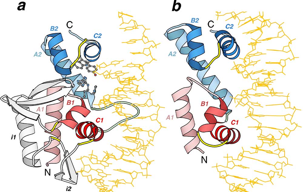
Structural similarity between Cre recombinase and MarA: Ribbon diagrams of a Cre recombinase from bacteriophage P1 (pdb entry 1crx, residues A154-A330) and b MarA transcription regulator from Escherichia coli (pdb entry 1bl0, residues A9-A106) in complex with DNA were drawn by Bobscript, a modified version of Molscript. The structures were superimposed and then separated for clarity. N- and C-termini are labeled. The spatially equivalent structural elements are colored correspondingly in the two structures. N-and C-terminal HHTH domains are colored red and blue respectively. α-Helices of the HTH motifs are in darker color. The turns in the HTH motifs are yellow and the loop connecting 2 HHTH domains is green. Long insertions (i1 and i2) in the first HHTH domain of Cre recombinase are shown in gray. DNA chains are orange. α-Helices are labeled as A, B, and C followed by a domain index (1 or 2). Side chains of active site residues in Cre-recombinase are shown in ball-and-stick presentation.