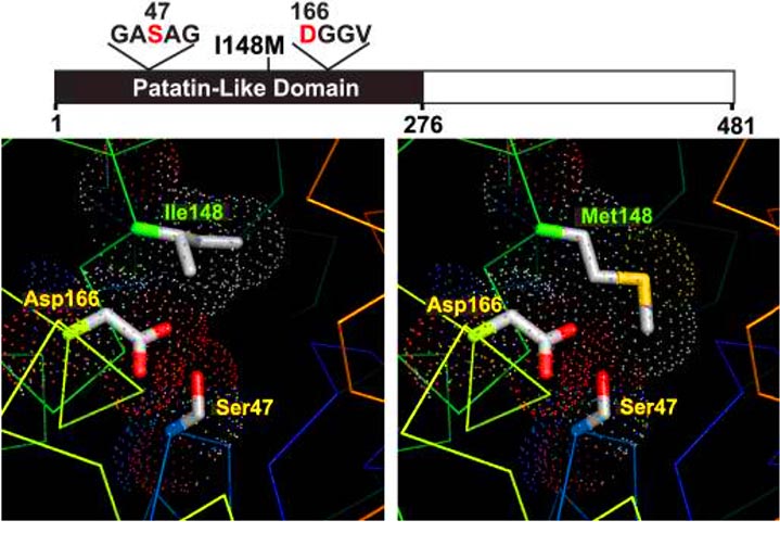
Structure models of wild type and mutant (I148M) PNPLA3. The domain structure of PNPLA3, showing the patatin-like domain (black) and locations of the catalytic dyad (Ser47 and Asp166) and the I148M substitution associated with increased hepatic triglyceride content, is shown. Structure models of normal (Ile148) and mutant (Met148) PNPLA3 are shown in the left and right panels, respectively. Protein traces are rainbow-colored from N to C terminus (blue to red) with side chains of catalytic dyad residues (positions 47 and 166) shown. The dots indicate a space-filling model corresponding to van der Waals atomic radii. Oxygen and sulfur atoms are colored red and yellow, respectively.