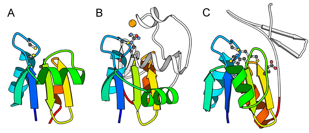
Thioredoxin-Like Folds. Ribbon diagrams represent (A) COG3019 rosetta model (DECOY_676), (B) bacterial disulfile oxidoreductase NrdH (1h75), and (C) E. coli arsenate reductase ArsC (1j9b). Corresponding secondary structural elements are colored identically in rainbow from the N-terminus to the C-terminus of the thioredoxin-like fold. Elements corresponding to inserted domains are white. Residues conserved between all three groups, which are involved in disulfide exchange, are depicted as a large red ball-and-stick. Residues conserved among individual groups are depicted as a ball-and-stick. The orange sphere in ArsC represents a sulfate ion and depicts the active site.