- A: few secondary structure elements
- X: Rubredoxin-like
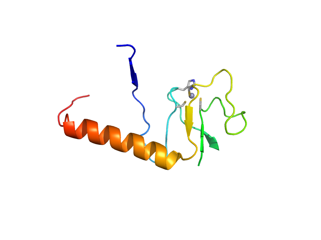
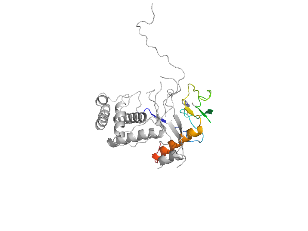
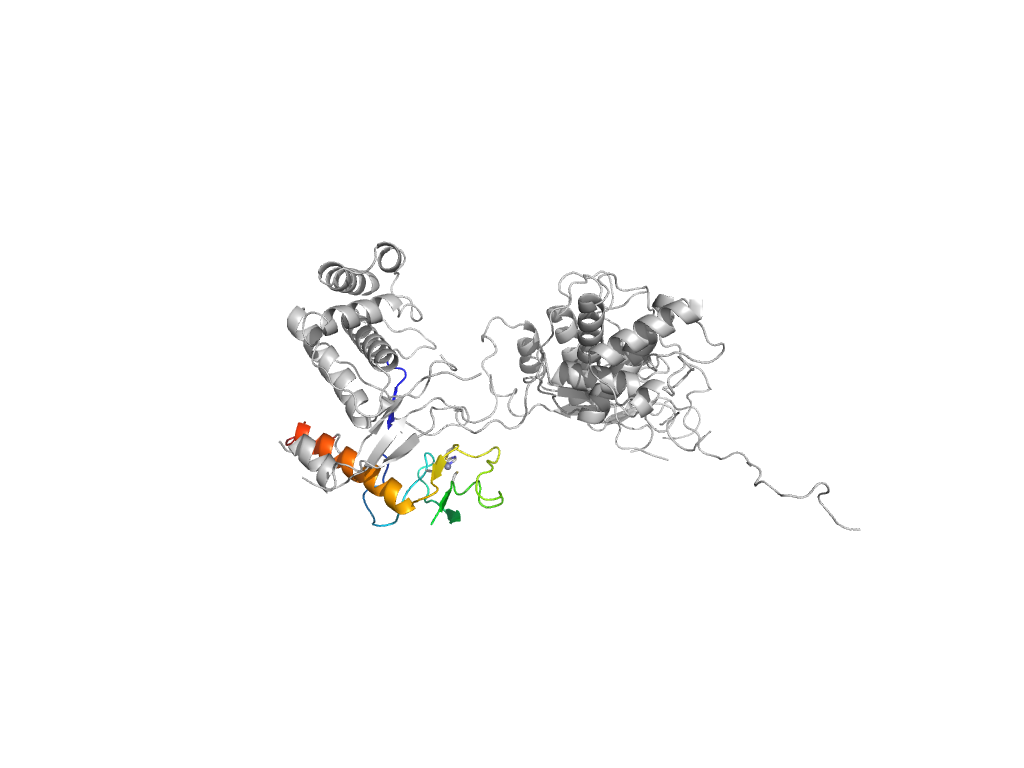
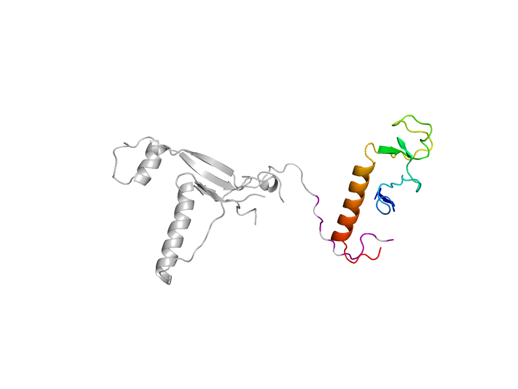
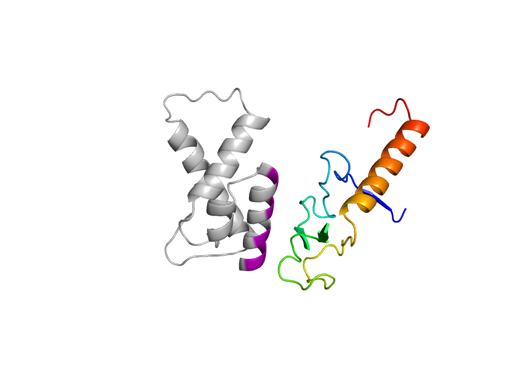
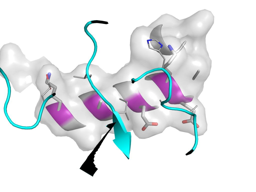
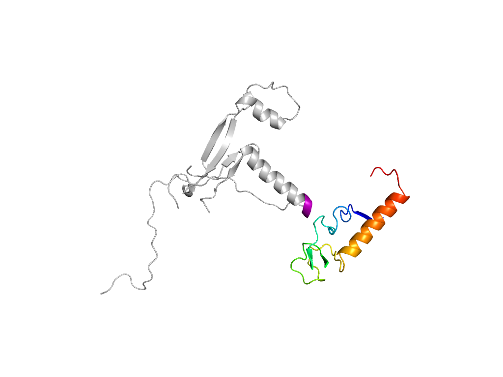
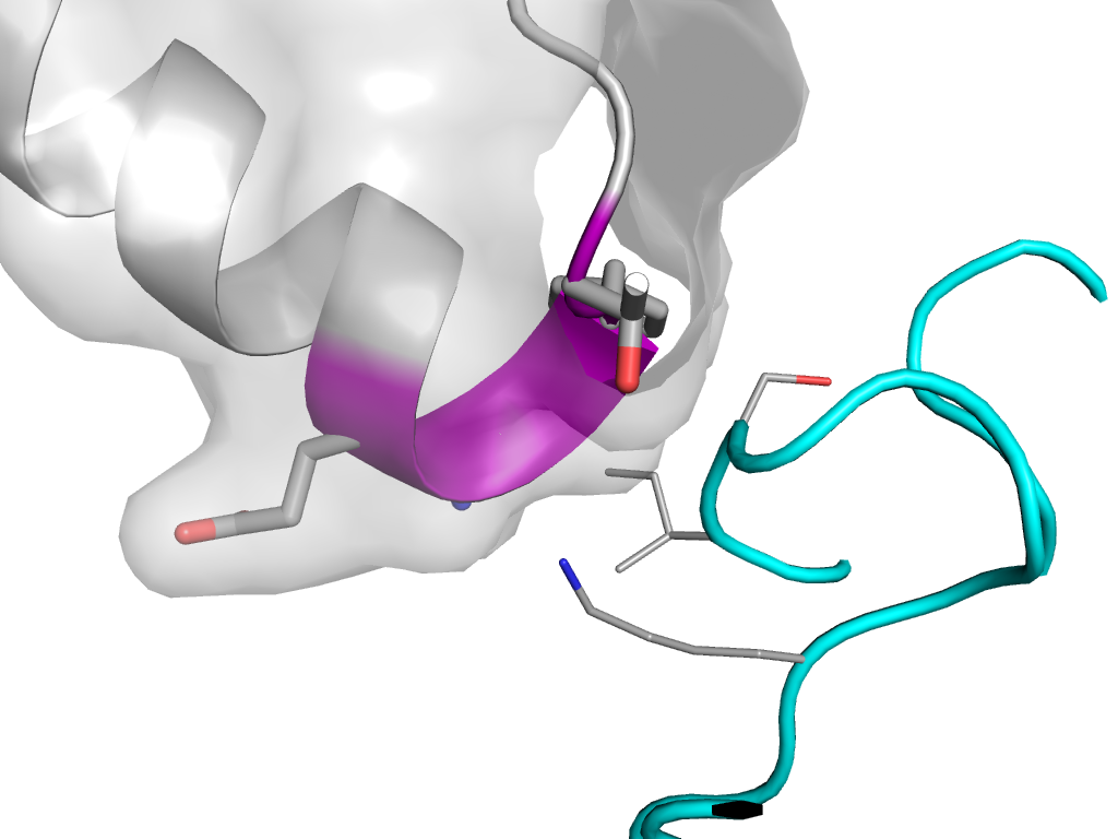
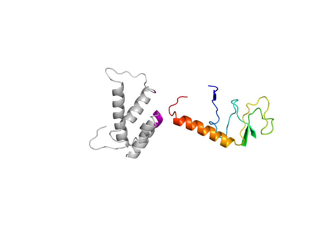
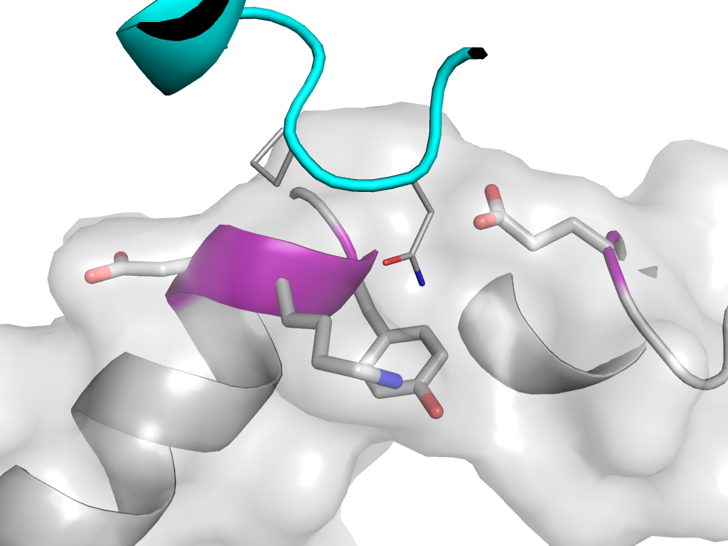
- H: Zn-binding domains of ADDBP
- T: Zn-binding domains of ADDBP
- F: Viral_DNA_Zn_bi
| UID: | 001117515 |
|---|---|
| Type: | Manual and representative domain |
| Range: | A:266-293,A:335-385 |
| Ligands: | ZN |
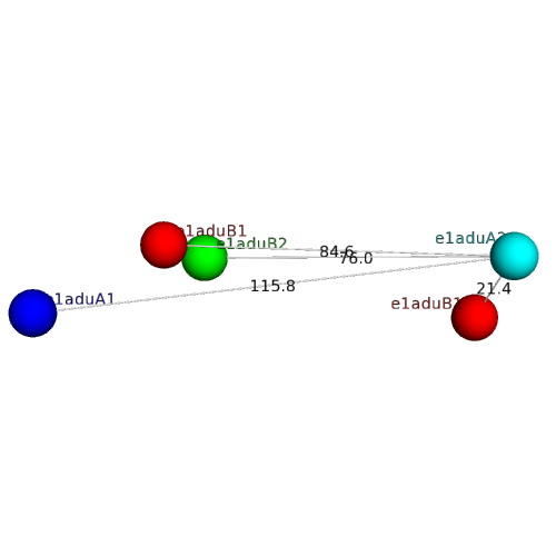
| PDB: | 1adu |
| PDB Description: | ADENOVIRUS SINGLE-STRANDED DNA-BINDING PROTEIN |
| UniProt: | P03265 |
| Species: |