- A: a/b three-layered sandwiches
- X: Periplasmic binding protein-like II
- H: Periplasmic binding protein-like II
- T: Periplasmic binding protein-like II
- F: Unmapped domains
| UID: | 001520877 |
|---|---|
| Type: | Automatic domain |
| Parent: | e1lstA3 |
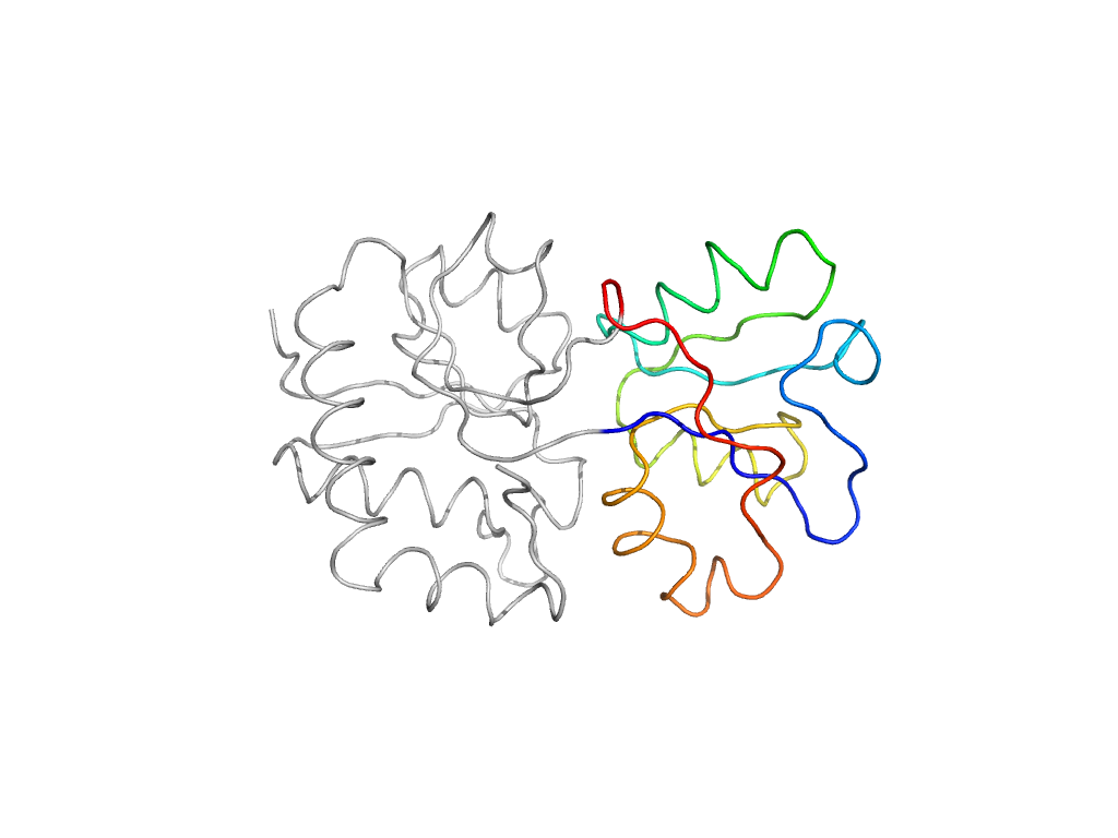
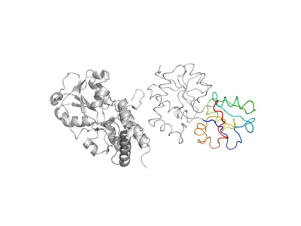
| Range: | B:91-190 |
| Ligands: | CD HIS |
| PDB: | 1hsl |
| PDB Description: | HISTIDINE-BINDING PROTEIN |
| UniProt: | P0AEU0 |
| Species: | Escherichia coli |

