| Domain ID | Symmetry operator | H-group | Visualization |
|---|
| e2fikB1 |
A:X,Y,Z->B:x,y,z |
Immunoglobulin-related |
Interaction
Interface
Pymol
|
| e2fikB1 |
A:-X+1,Y-1/2,-Z+1/2->B:x,y,z |
Immunoglobulin-related |
Interaction
Interface
Pymol
|
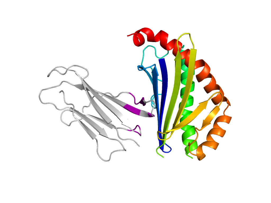
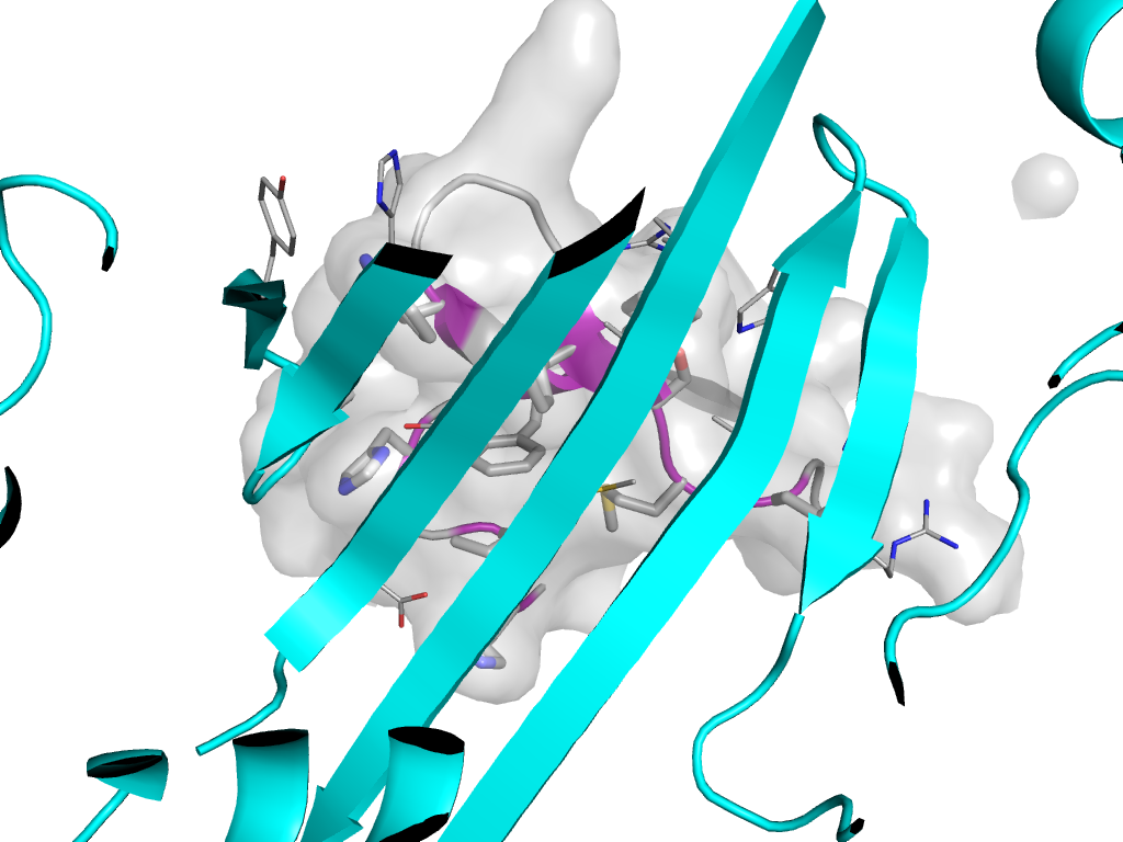
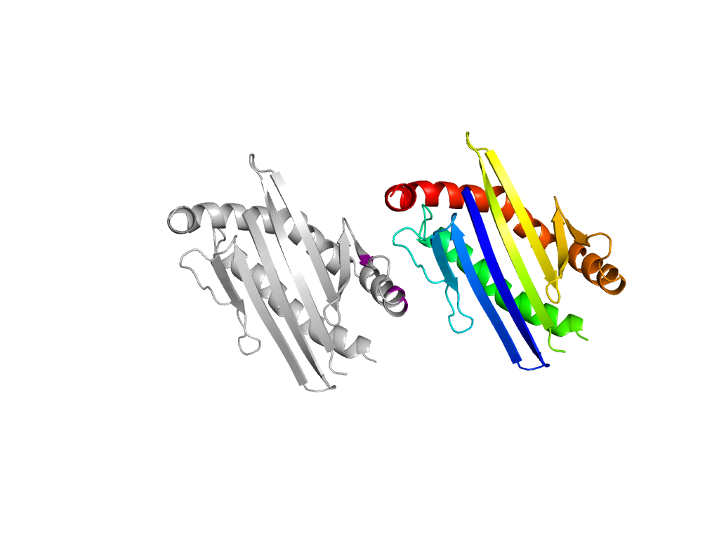
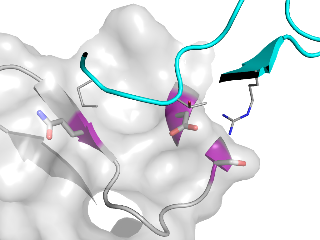
| e2fikA2 |
A:x,y,z->A:X-1,Y,Z |
MHC antigen-recognition domain |
Interaction
Interface
Pymol
|
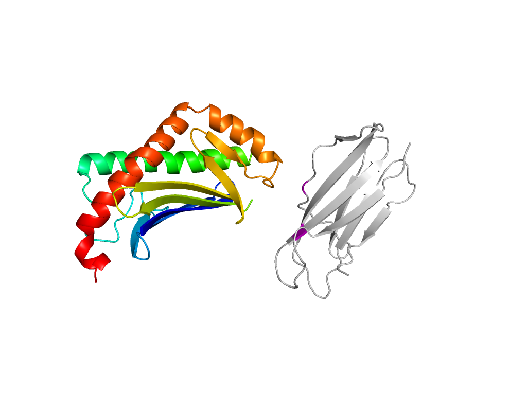
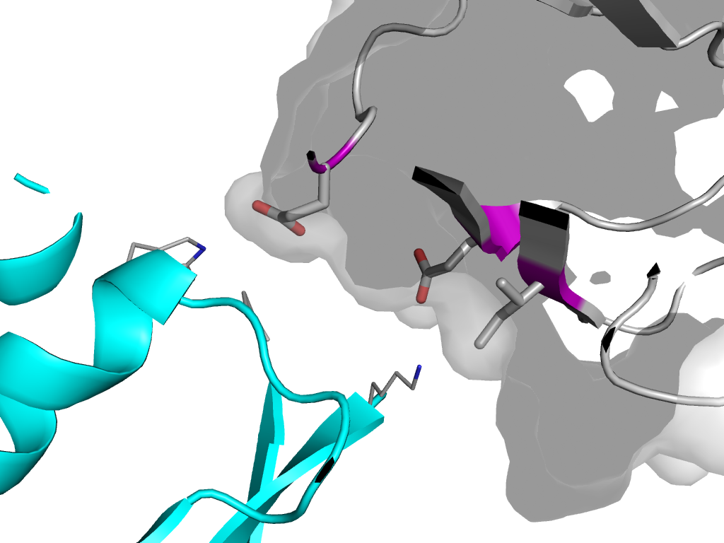
| e2fikA2 |
A:X-1,Y,Z->A:x,y,z |
MHC antigen-recognition domain |
Interaction
Interface
Pymol
|
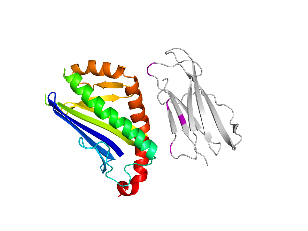
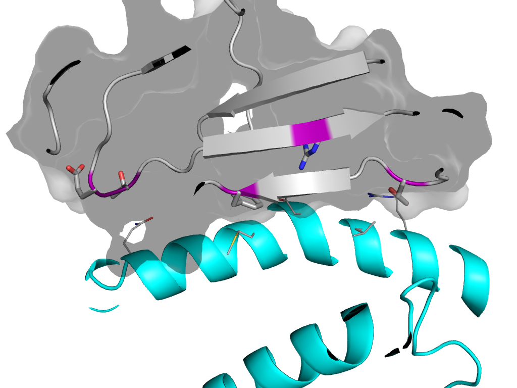
| e2fikA1 |
A:X-1,Y,Z->A:x,y,z |
Immunoglobulin-related |
Interaction
Interface
Pymol
|
| e2fikB1 |
A:X-1,Y,Z->B:x,y,z |
Immunoglobulin-related |
Interaction
Interface
Pymol
|
| e2fikA1 |
A:-X+1,Y-1/2,-Z+1/2->A:x,y,z |
Immunoglobulin-related |
Interaction
Interface
Pymol
|