Domain ID e2ihoA1

Download files:
  • A: beta duplicates or obligate multimers
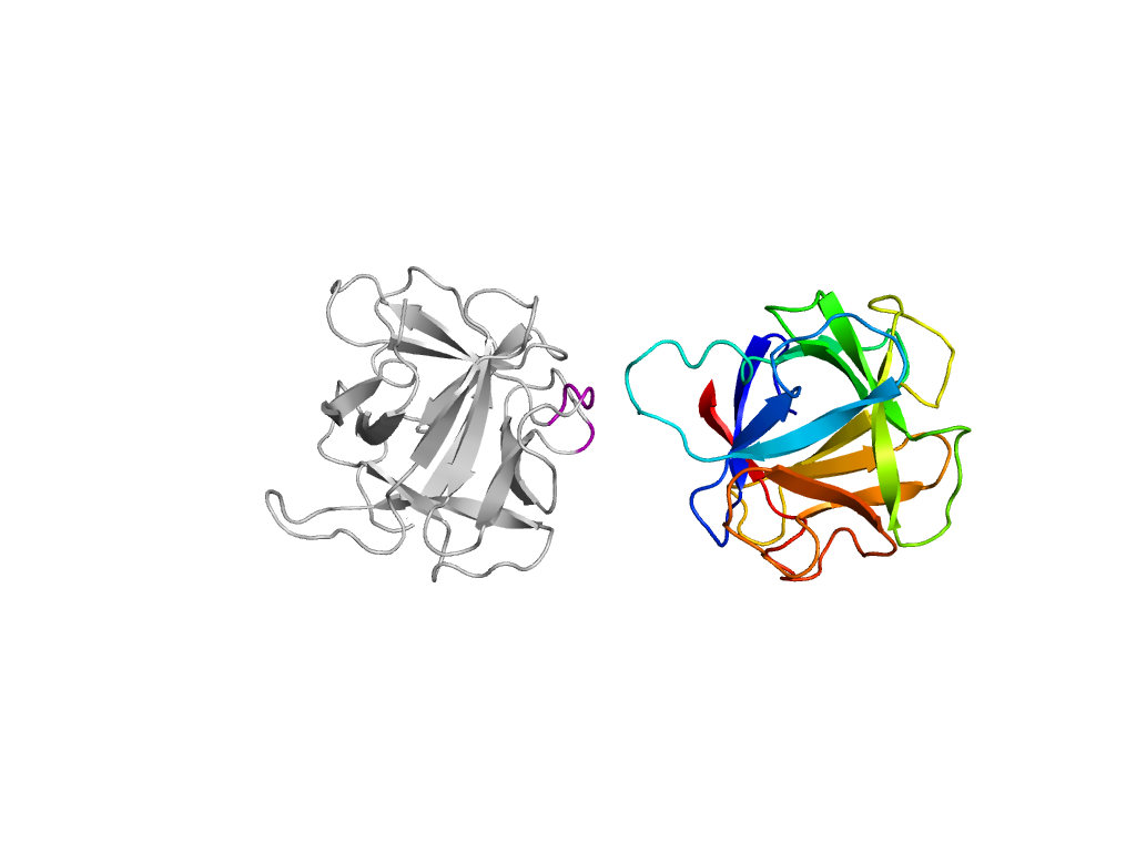
  • X: beta-Trefoil
  • H: beta-Trefoil
  • T: beta-Trefoil
  • F: RicinB_lectin_2
UID: 000002130
Type: Manual and representative domain
Range: A:2-155
PDB: 2iho
PDB Description: Lectin
UniProt: Q8X123
Species: Marasmius oreades

Structure of domain e2ihoA1

Structural contacts of domain e2ihoA1 of H-group "beta-Trefoil":

The domain and its neighboring domains (both within the asymmetric unit and to crystallographic symmetry mates) are represented by spheres and linked by lines. Distances between the center of the domain and interfaces are shown for each contact.

Domain IDSymmetry operatorH-groupVisualization
e2ihoA1 A:x,y,z->A:-Y+1,X-Y,Z-1/3 beta-Trefoil Interaction Interface Pymol
e2ihoA2 A:-Y+1,X-Y,Z-1/3->A:x,y,z Cysteine proteinases Interaction Interface Pymol
e2ihoA1 A:-Y+1,X-Y,Z-1/3->A:x,y,z beta-Trefoil Interaction Interface Pymol