- A: beta duplicates or obligate multimers
- X: Triple beta-spiral
- H: Triple beta-spiral
- T: Triple beta-spiral
- F: Reo_sigmaC_M
| UID: | 001833577 |
|---|---|
| Type: | Automatic and representative domain |
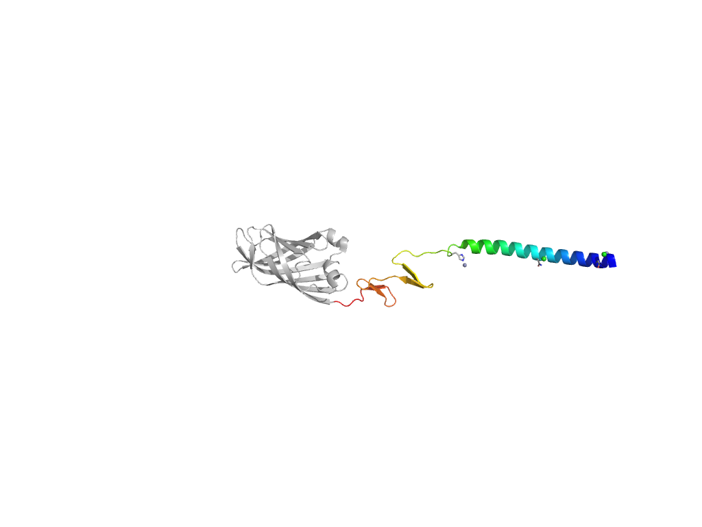
| Parent: | e2vrsC1 |
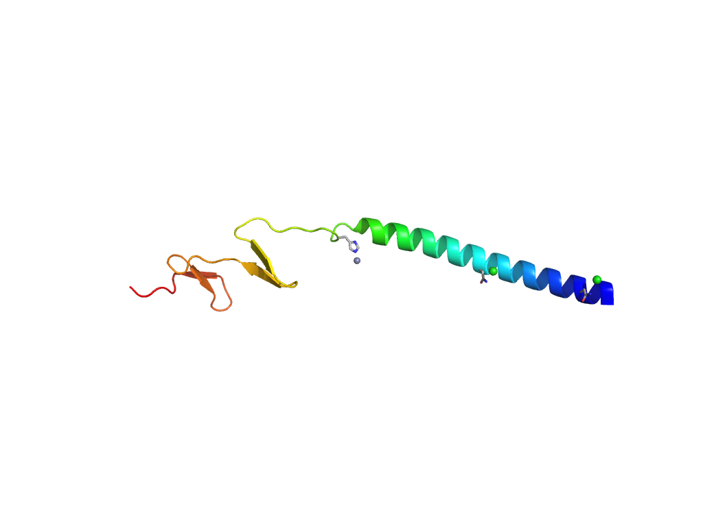
| Range: | A:120-195 |
| Ligands: | CL ZN |
| PDB: | 2jjl |
| PDB Description: | SIGMA-C CAPSID PROTEIN |
| UniProt: | Q992I2 |
| Species: | Avian orthoreovirus |

