- A: a/b three-layered sandwiches
- X: Periplasmic binding protein-like II
- H: Periplasmic binding protein-like II
- T: Periplasmic binding protein-like II
- F: VitK2_biosynth
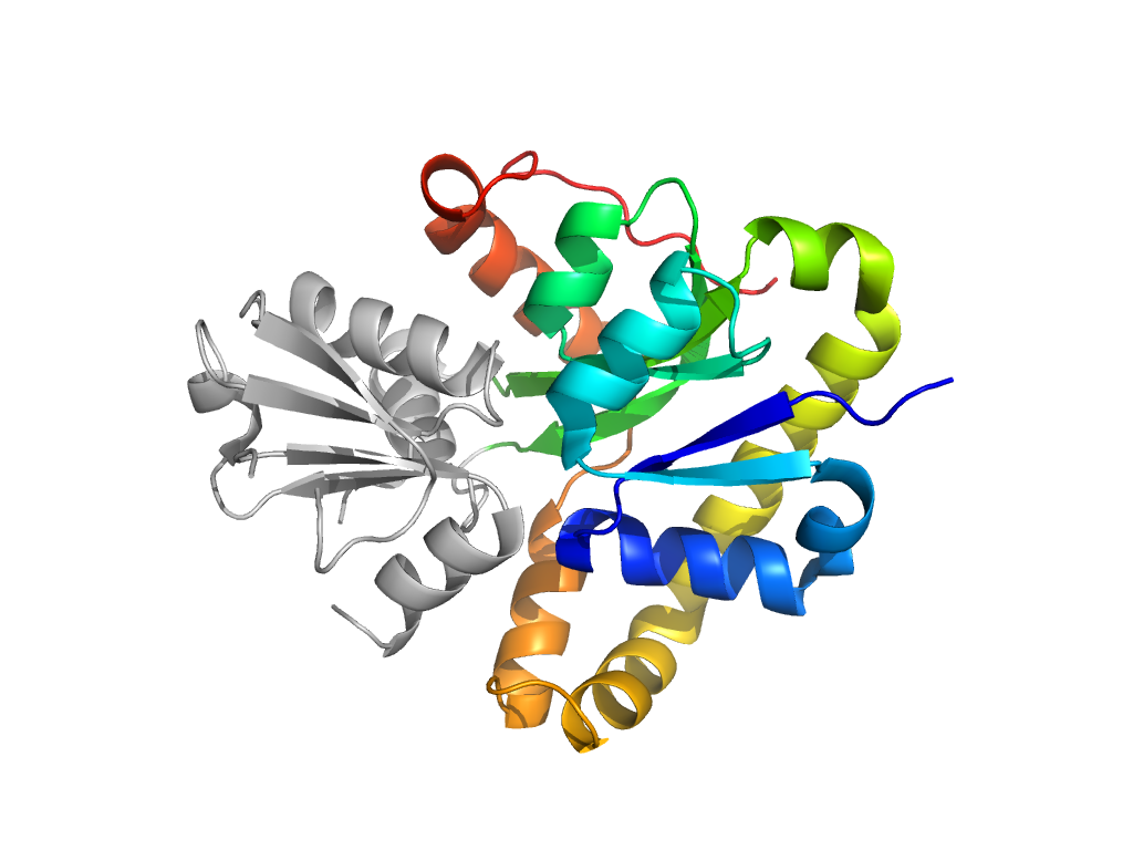
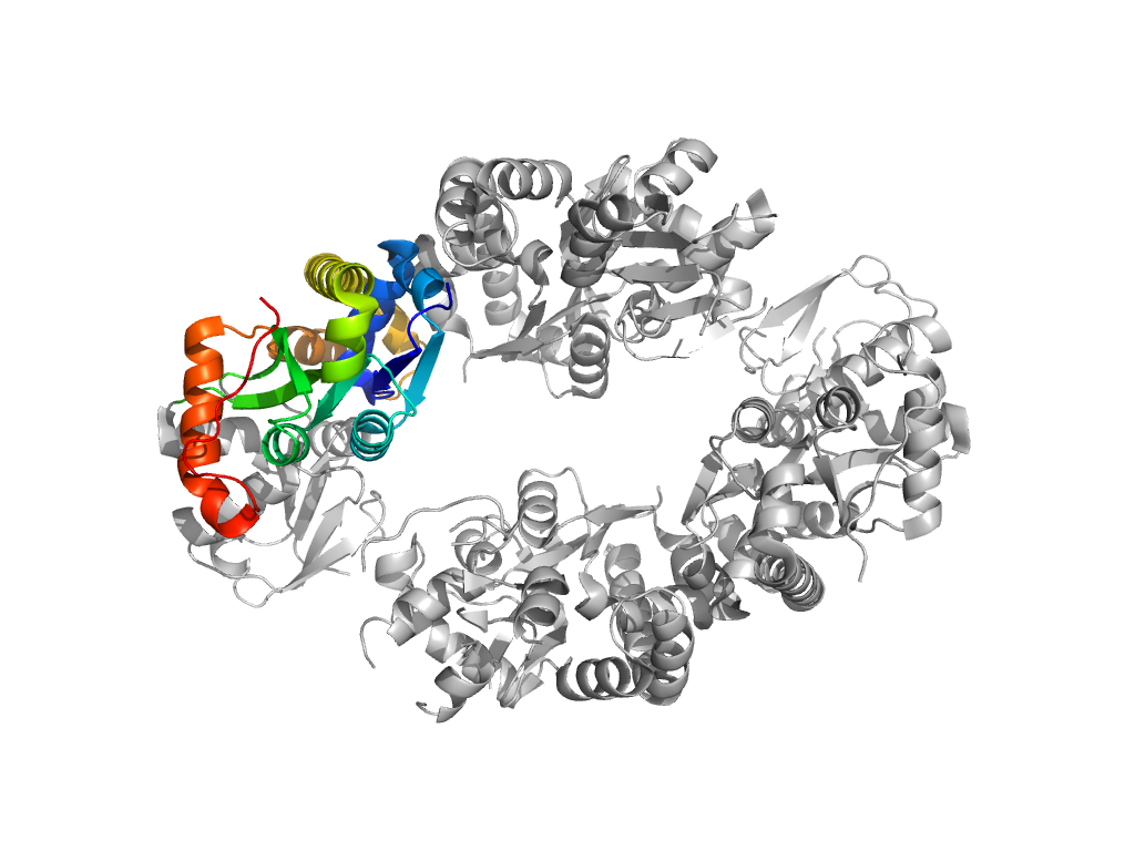
| UID: | 001521878 |
|---|---|
| Type: | Automatic domain |
| Parent: | e2nxoA2 |
| Range: | C:4-80,C:181-280 |
| PDB: | 2nxo |
| PDB Description: | Hypothetical protein SCO4506 |
| UniProt: | Q9L0T8 |
| Species: | Streptomyces coelicolor |

