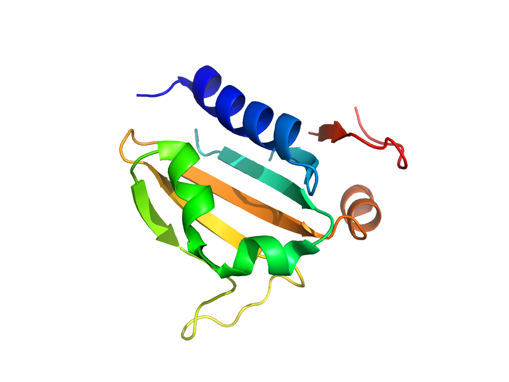
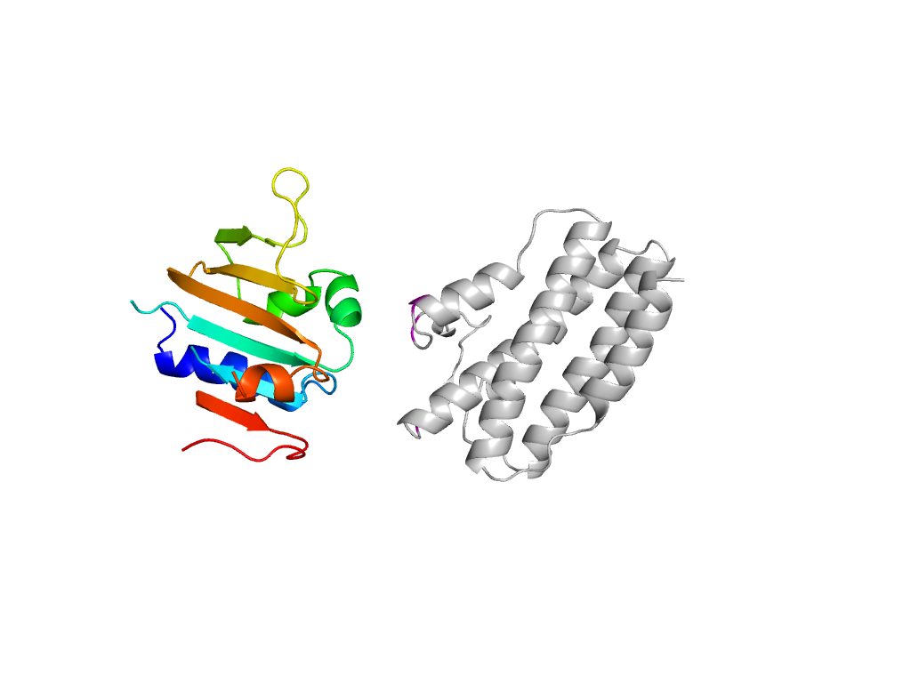
- A: a+b three layers
- X: Nucleotidyltransferase-like
- H: Nucleotidyltransferase
- T: Nucleotidyltransferase
- F: Adenyl_transf
| UID: | 000006831 |
|---|---|
| Type: | Manual and representative domain |
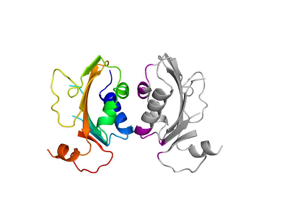
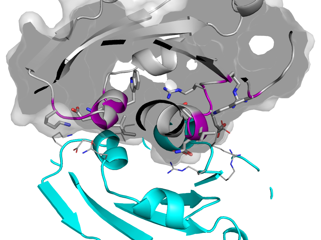
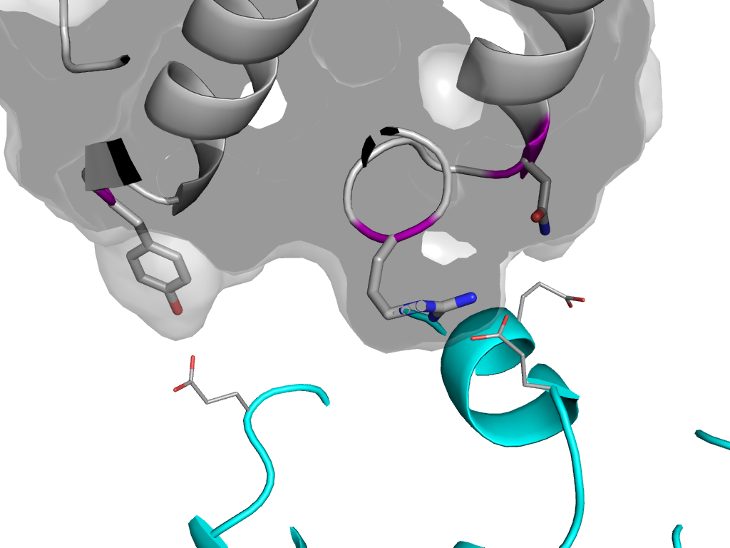
| Range: | A:1-135 |
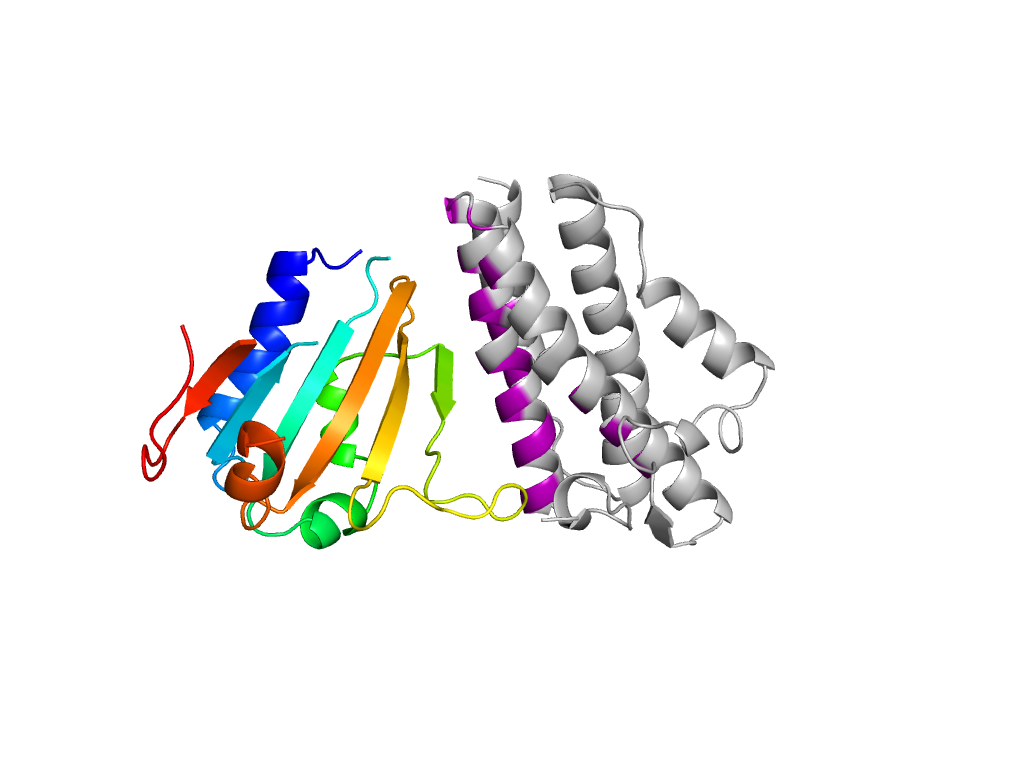
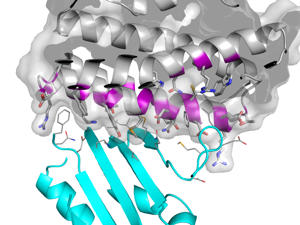
| PDB: | 2pbe |
| PDB Description: | Aminoglycoside 6-adenylyltransferase |
| UniProt: | P17585 |
| Species: | Bacillus subtilis |

