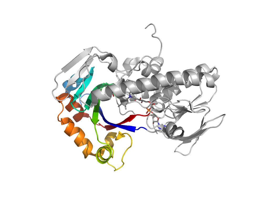
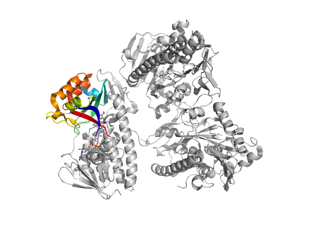
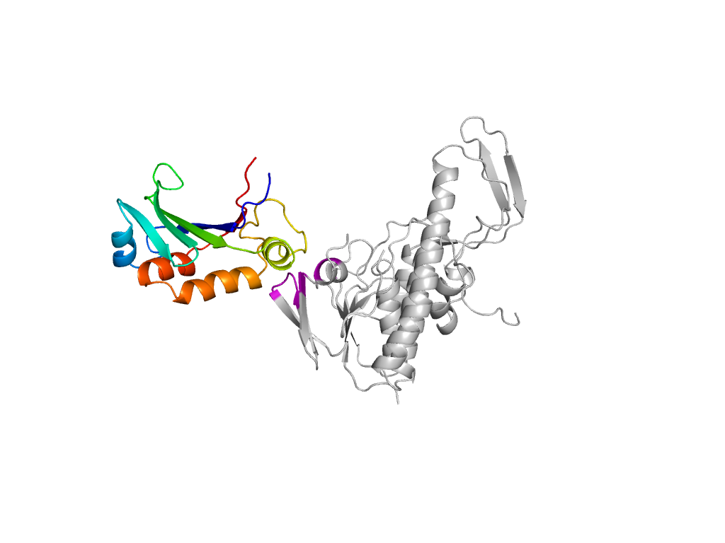
- A: a+b two layers
- X: FAD-linked reductases, C-terminal domain-like
- H: FAD-linked reductases-C
- T: FAD-linked reductases-C
- F: FAD_binding-like
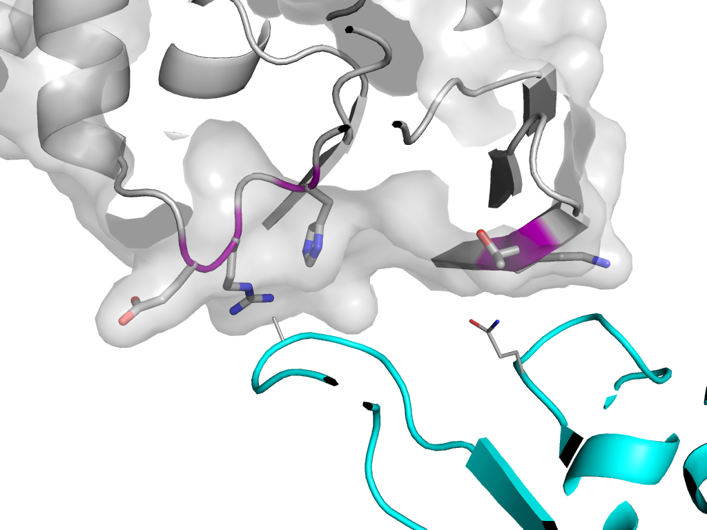
| UID: | 000006698 |
|---|---|
| Type: | Manual and representative domain |
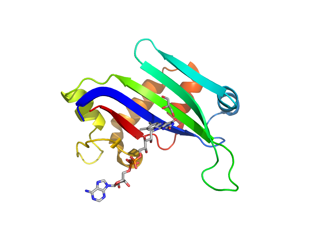
| Range: | A:164-291 |
| Ligands: | FAD GOL |
| PDB: | 2vou |
| PDB Description: | 2,6-DIHYDROXYPYRIDINE HYDROXYLASE |
| UniProt: | Q93NG3 |
| Species: | Arthrobacter nicotinovorans |
