- A: alpha arrays
- X: 60S acidic ribosomal protein P1/P2
- H: 60S acidic ribosomal protein P1/P2
- T: 60S acidic ribosomal protein P1/P2
- F: Ribosomal_60s
| UID: | 001840982 |
|---|---|
| Type: | Manual and representative domain |
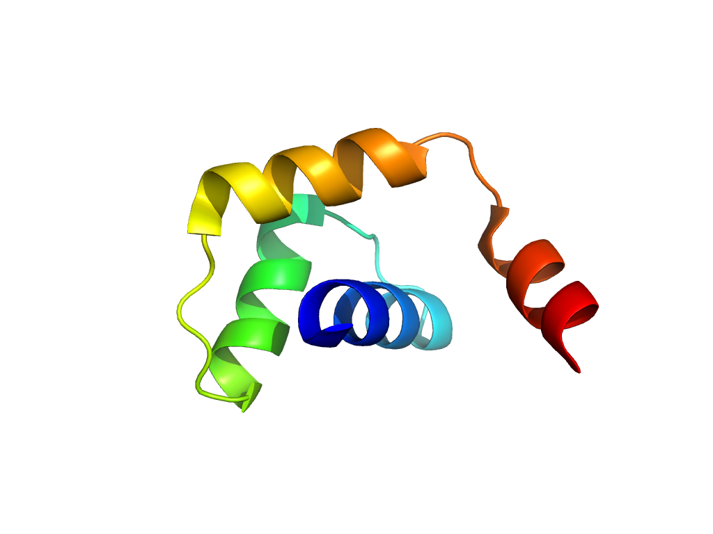
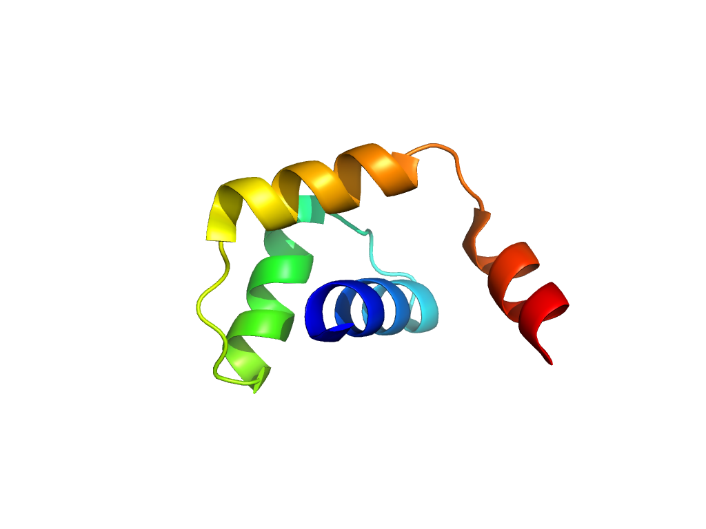
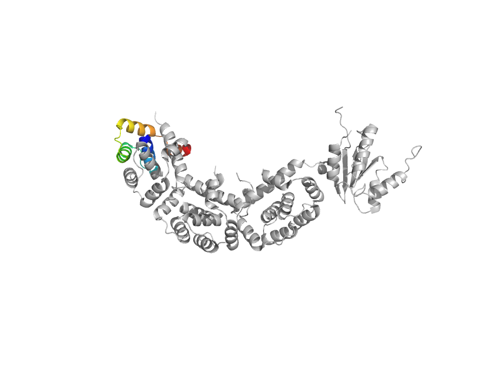
| Range: | A:1-58 |
| PDB: | 3a1y |
| PDB Description: | 50S ribosomal protein P1 (L12P) |
| UniProt: | O57705 |
| Species: | Pyrococcus horikoshii |
