| UID: | 000414844 |
|---|---|
| Type: | Automatic domain |
| Parent: | e1cxqA1 |
| Range: | B:57-209 |
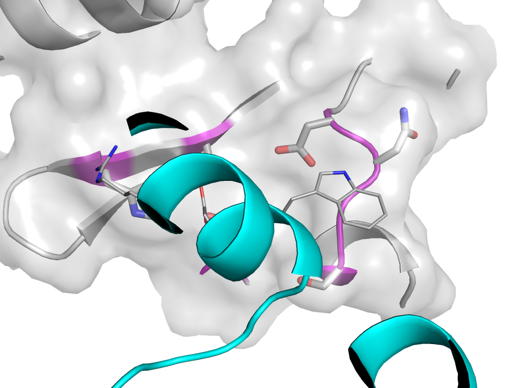
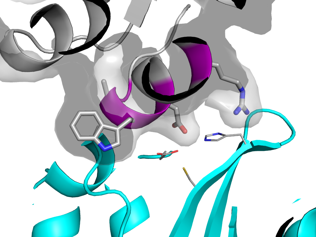
| Ligands: | AVX CD CL DTT SO4 |
| PDB: | 3ao2 |
| PDB Description: | POL POLYPROTEIN |
| UniProt: | P04585 |
| Species: | Human immunodeficiency virus 1 |
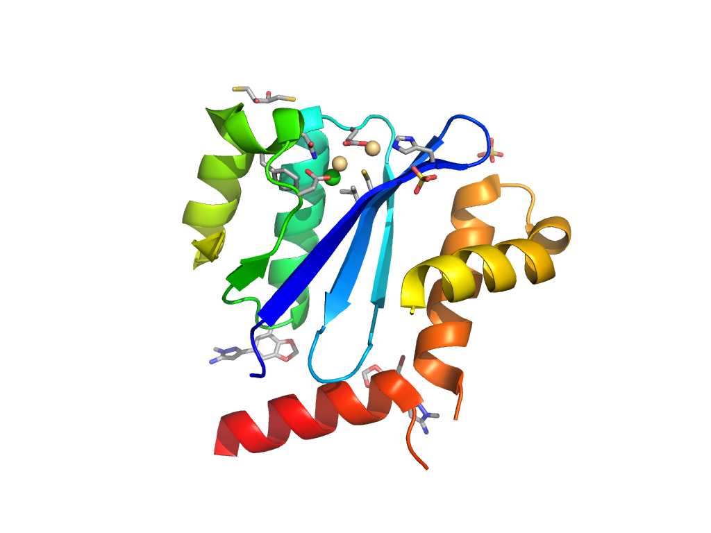
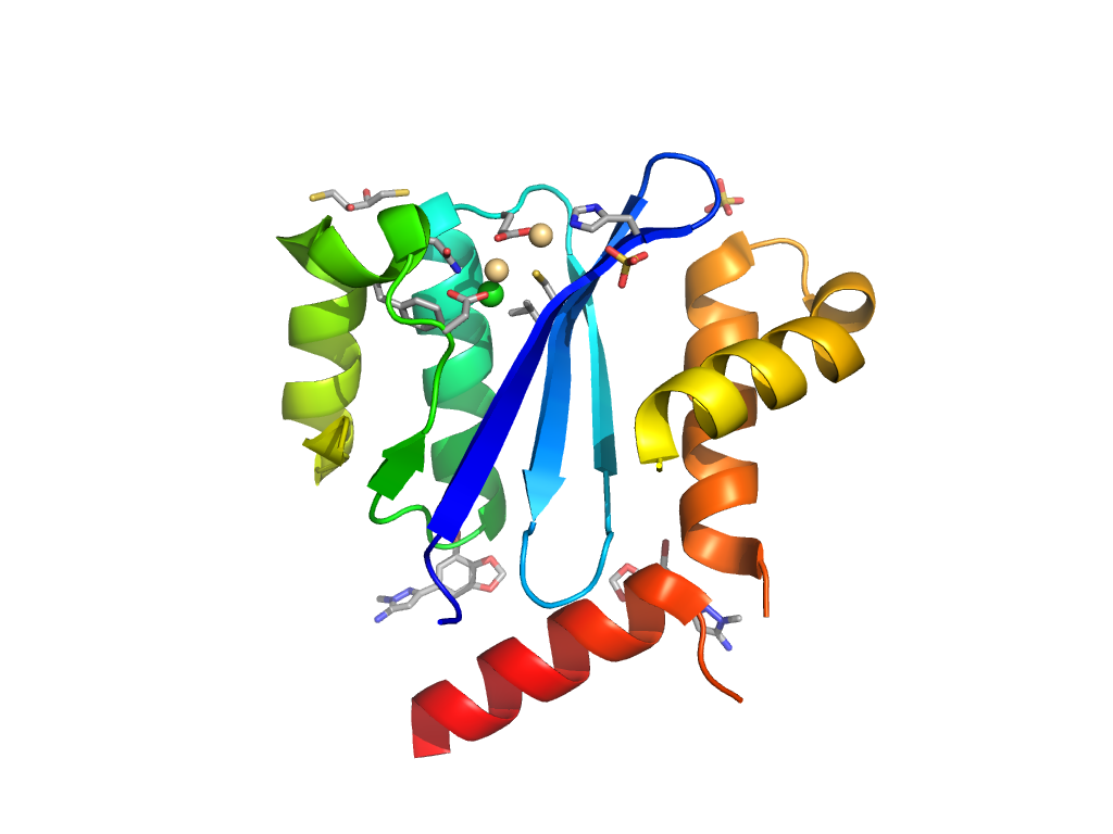
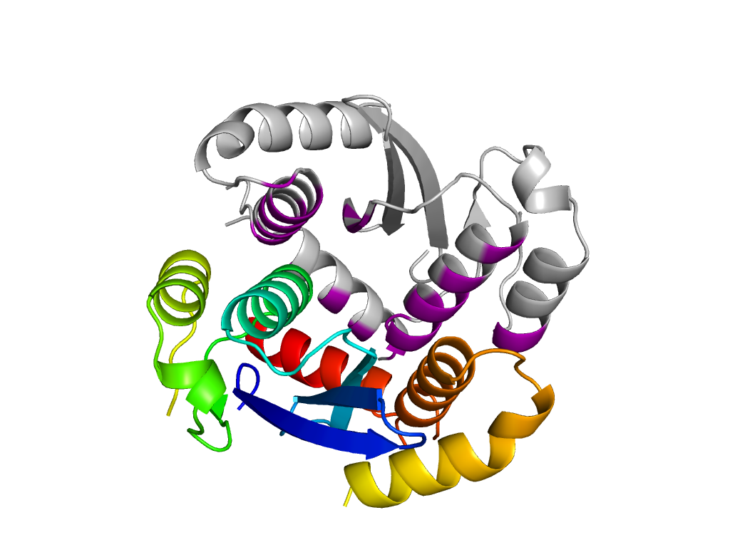
Structure of domain e3ao2B1
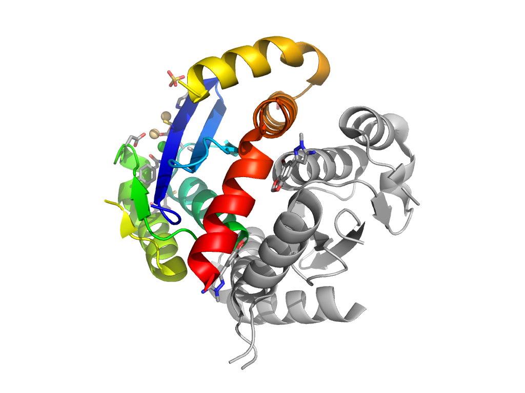
Domains in the same chain:
| e3ao2B1 | B:57-209 | Ribonuclease H-like | Ribonuclease H-like | Ribonuclease H-like |
Domains in the same PDB:
| e3ao2A1 | A:57-209 | Ribonuclease H-like | Ribonuclease H-like | Ribonuclease H-like |
| e3ao2B1 | B:57-209 | Ribonuclease H-like | Ribonuclease H-like | Ribonuclease H-like |
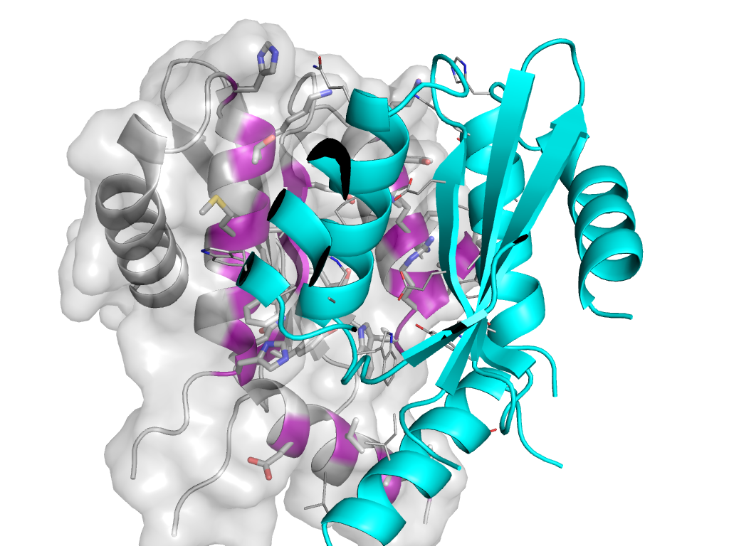
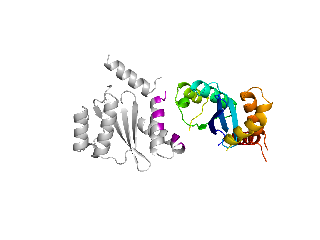
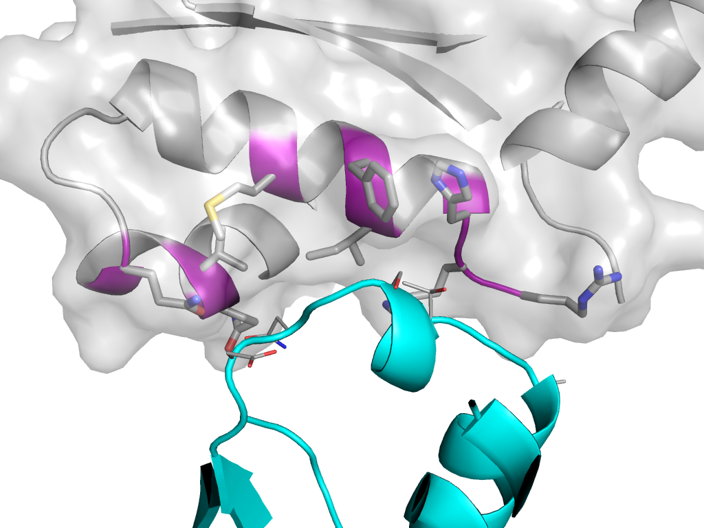
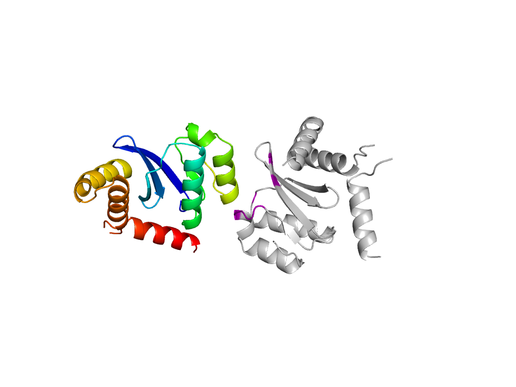
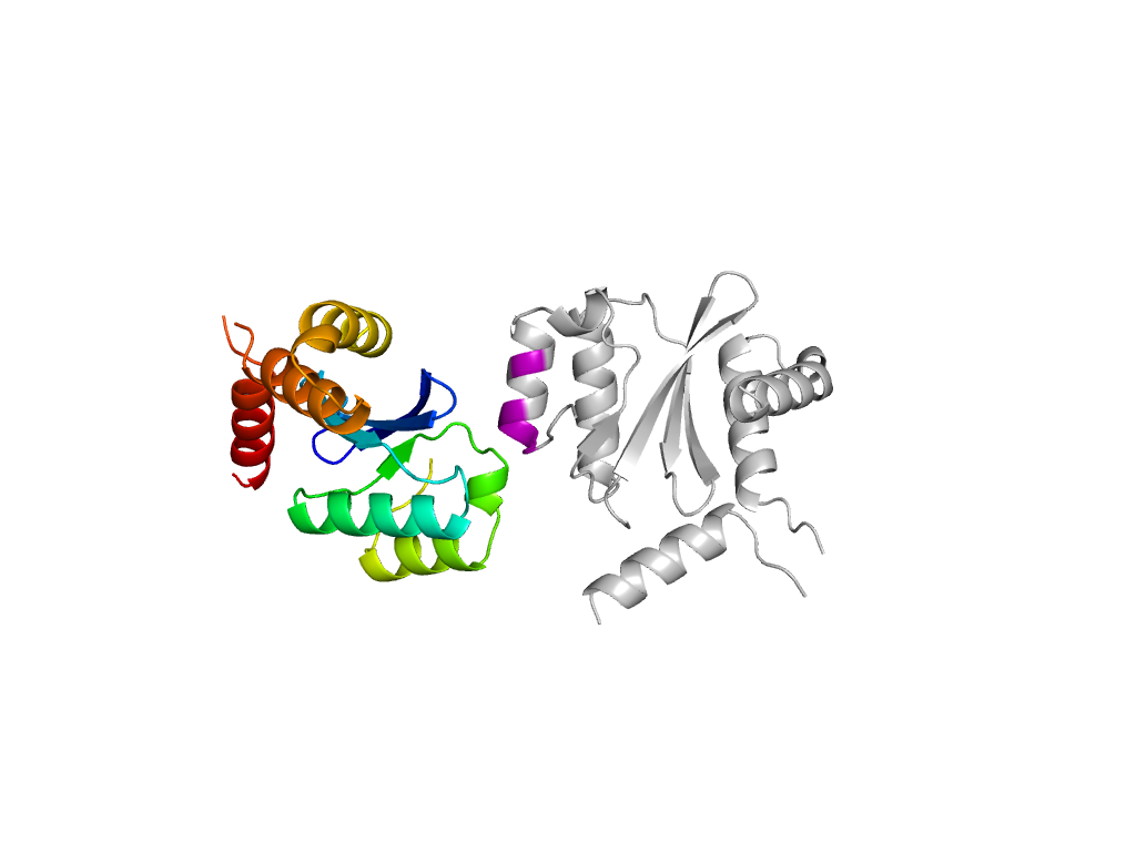
Structural contacts of domain e3ao2B1 of H-group "Ribonuclease H-like":
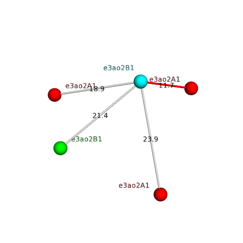
The domain and its neighboring domains (both within the asymmetric unit and to crystallographic symmetry mates) are represented by spheres and linked by lines. Distances between the center of the domain and interfaces are shown for each contact.
| Domain ID | Symmetry operator | H-group | Visualization |
|---|---|---|---|
| e3ao2A1 | B:x,y,z->A:X,Y,Z | Ribonuclease H-like | Interaction Interface Pymol |
| e3ao2B1 | B:x,y,z->B:-Y,X-Y,Z-1/3 | Ribonuclease H-like | Interaction Interface Pymol |
| e3ao2B1 | B:-Y,X-Y,Z-1/3->B:x,y,z | Ribonuclease H-like | Interaction Interface Pymol |
| e3ao2A1 | B:x,y,z->A:-Y,X-Y-1,Z-1/3 | Ribonuclease H-like | Interaction Interface Pymol |
| e3ao2A1 | B:x,y,z->A:-Y,X-Y,Z-1/3 | Ribonuclease H-like | Interaction Interface Pymol |