- A: a/b three-layered sandwiches
- X: Periplasmic binding protein-like II
- H: Periplasmic binding protein-like II
- T: Periplasmic binding protein-like II
- F: SBP_bac_11
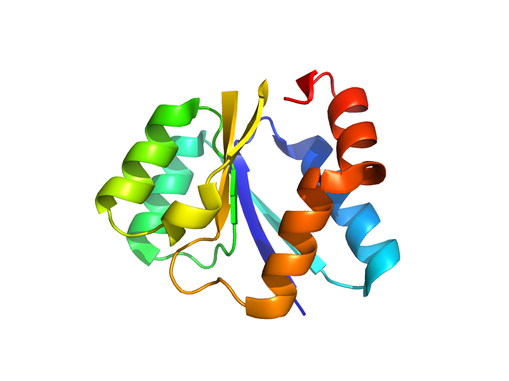
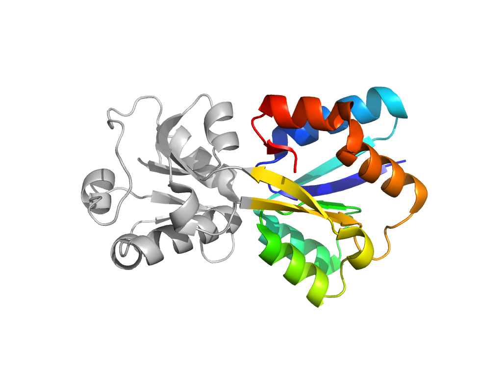
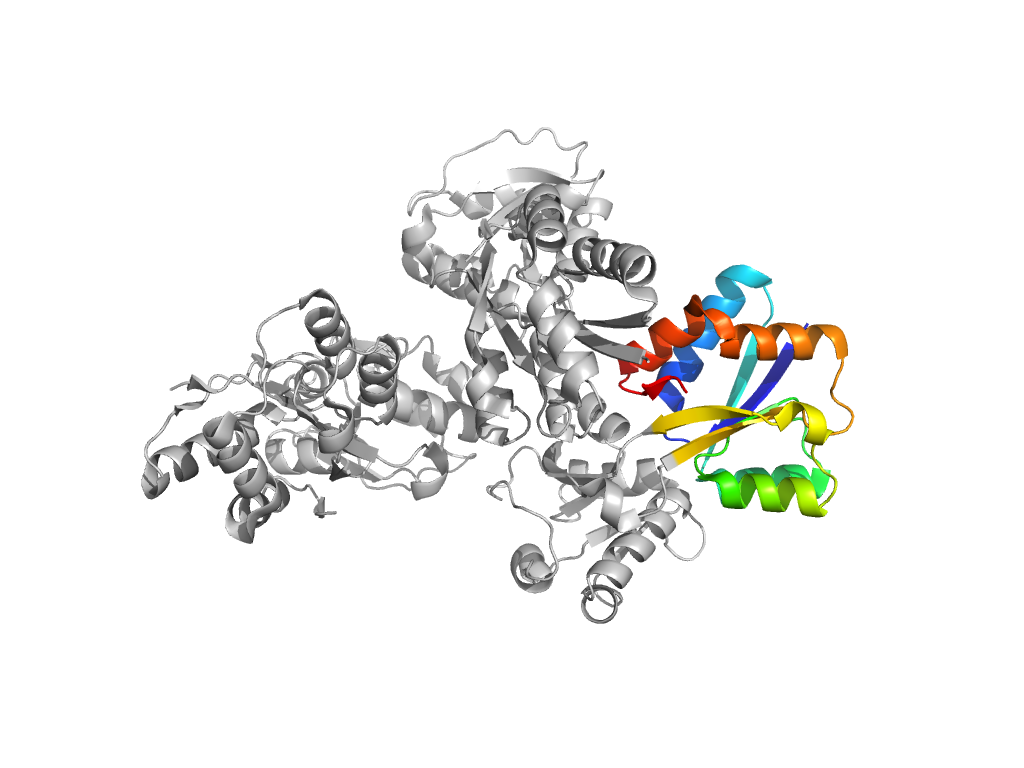
| UID: | 001519542 |
|---|---|
| Type: | Automatic domain |
| Parent: | e1amfA2 |
| Range: | B:3-82,B:195-233 |
| Ligands: | REO |
| PDB: | 3axf |
| PDB Description: | MOLYBDATE-BINDING PERIPLASMIC PROTEIN |
| UniProt: | P37329 |
| Species: |
