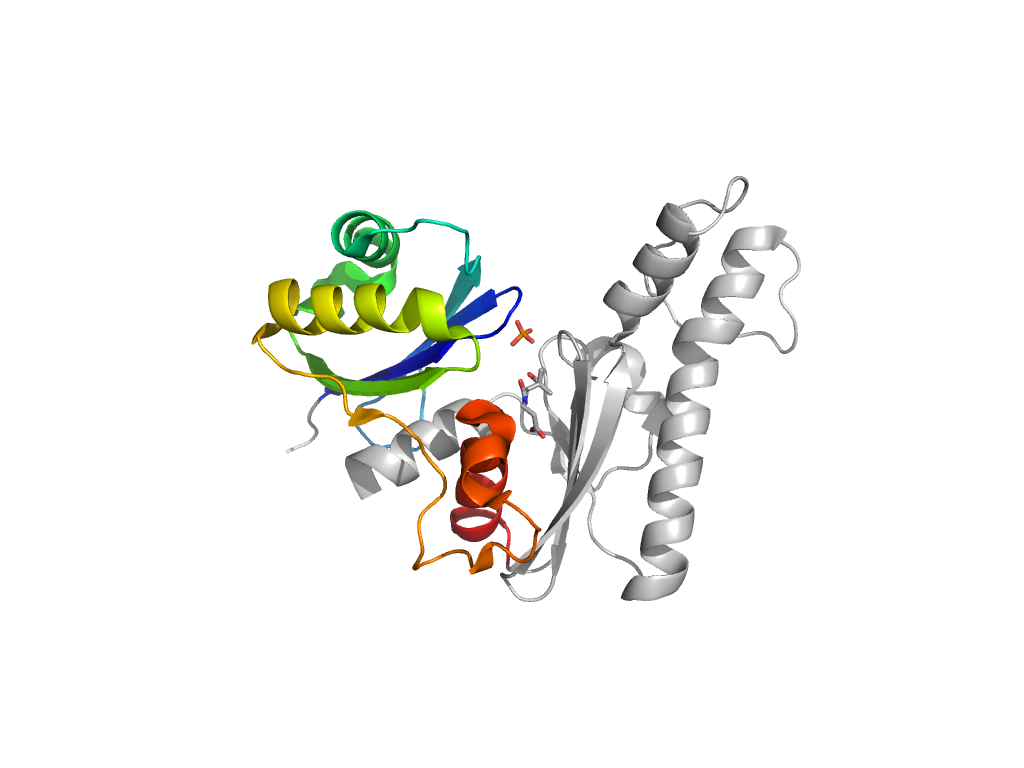
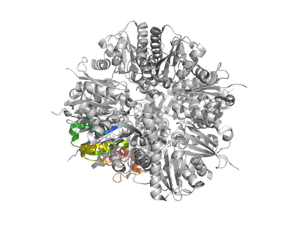
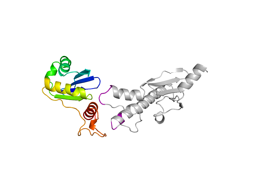
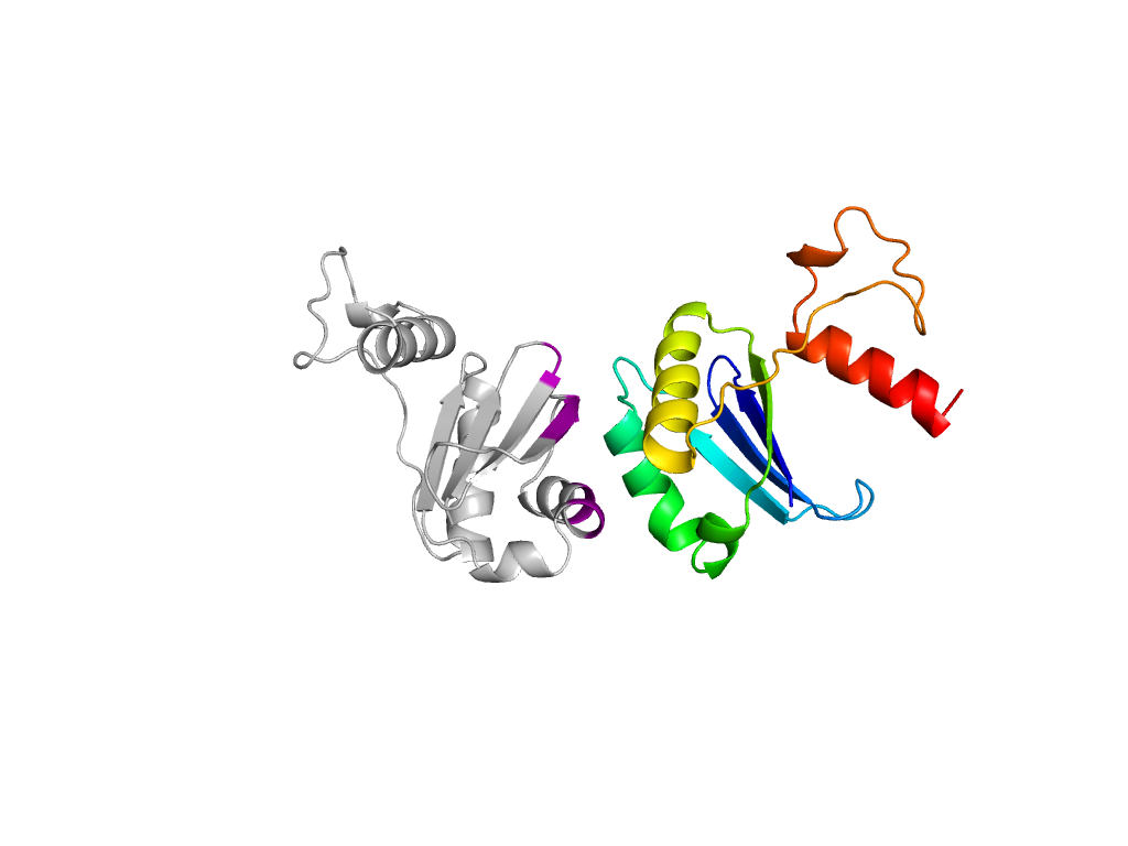
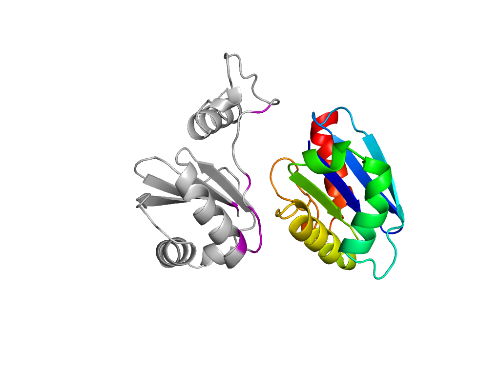
- A: mixed a+b and a/b
- X: Ribonuclease H-like
- H: Ribonuclease H-like
- T: Ribonuclease H-like
- F: Pan_kinase
| UID: | 000011231 |
|---|---|
| Type: | Manual and representative domain |
| Range: | A:1-118 |
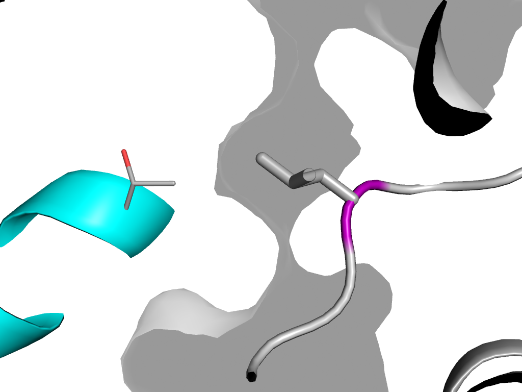
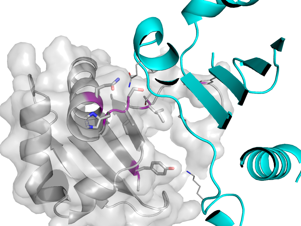
| Ligands: | PAU PO4 |
| PDB: | 3bex |
| PDB Description: | TYPE III PANTOTHENATE KINASE |
| UniProt: | Q9WZY5 |
| Species: |

