Domain ID e3bz1B2

Download files:
  • A: a+b two layers
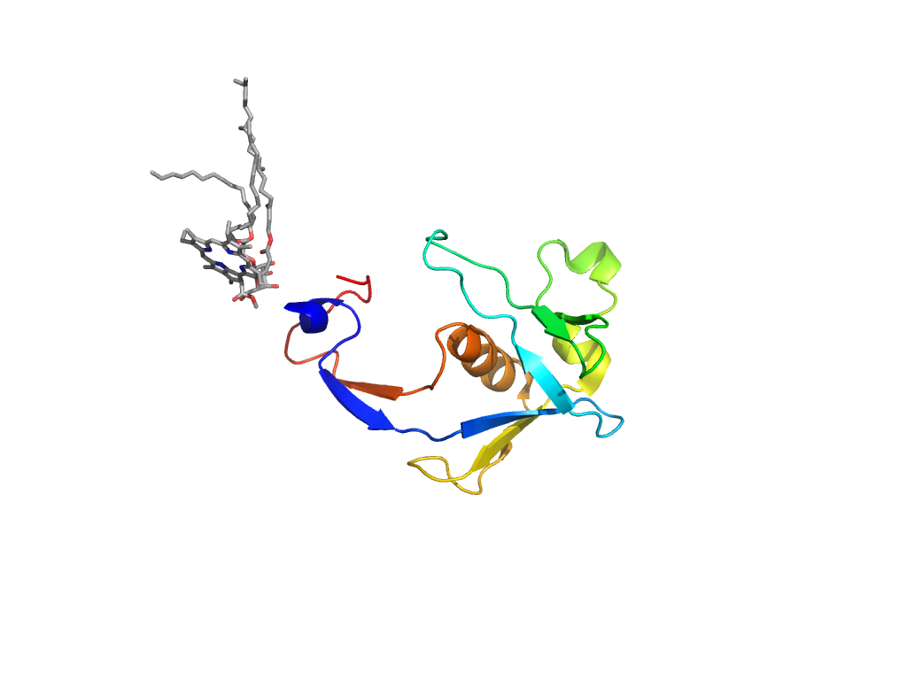
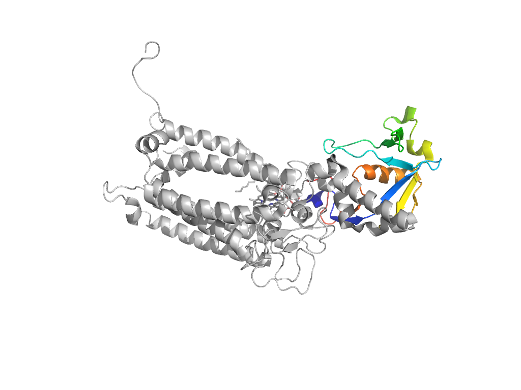
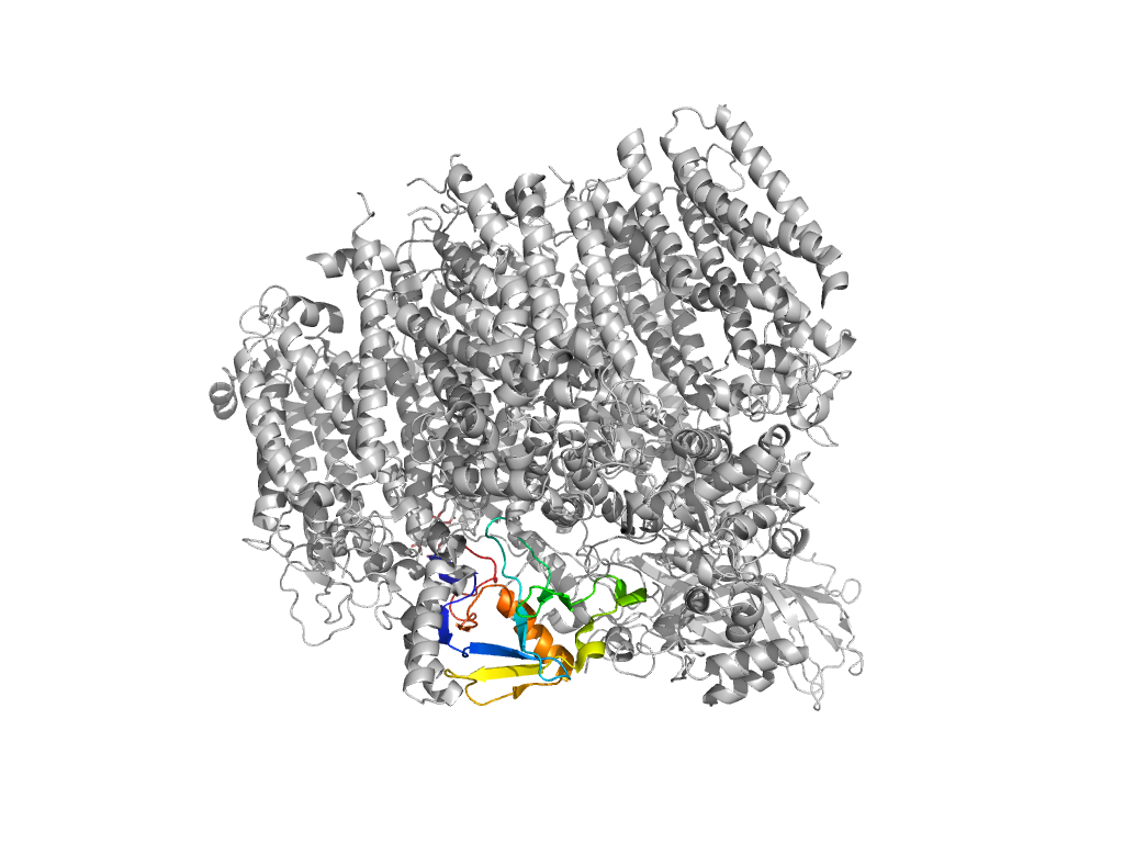
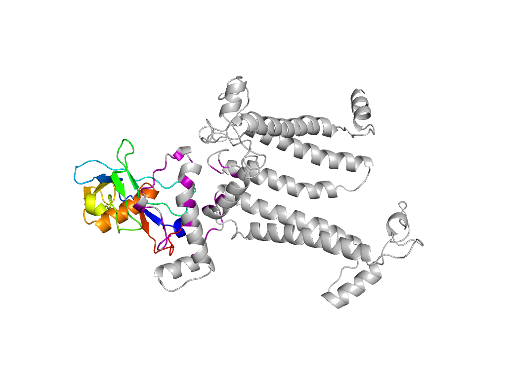
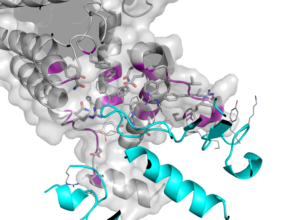
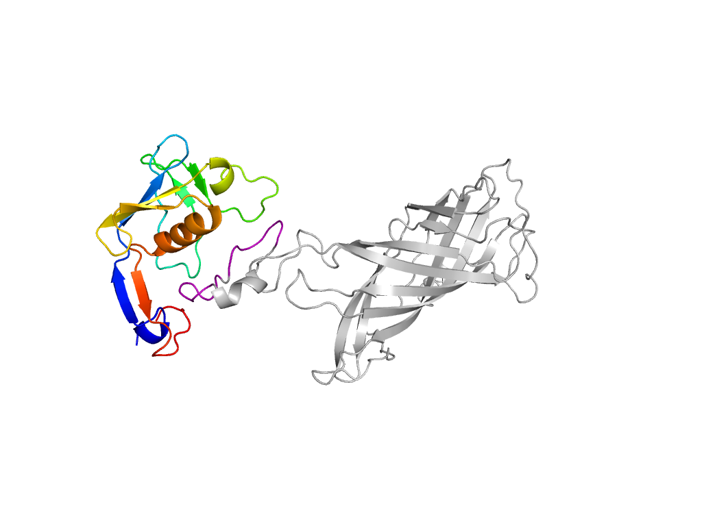
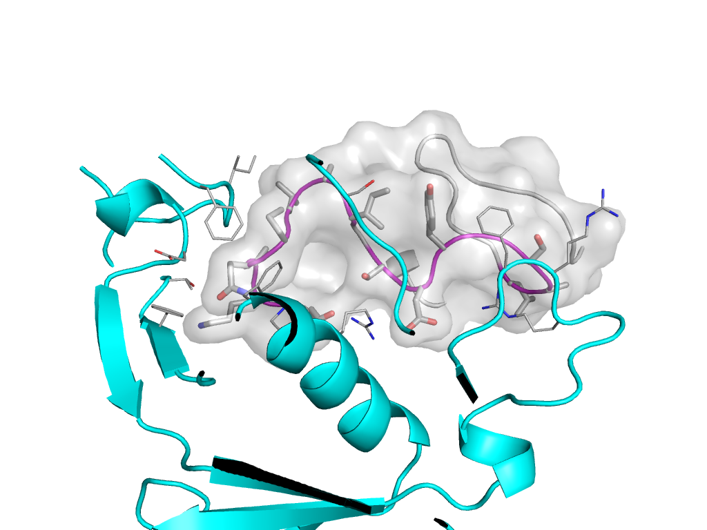
  • X: Photosystem II antenna protein PsbB insertion domain
  • H: Photosystem II antenna protein PsbB insertion domain
  • T: Photosystem II antenna protein PsbB insertion domain
  • F: PSII
UID: 001108422
Type: Automatic domain
Parent: e2axtB3
Range: B:329-444
Ligands: CLA LMG
PDB: 3bz1
PDB Description:
Species:

Structure of domain e3bz1B2

Domains in the same chain:

e3bz1B1 B:2-328,B:445-491 Photosystem II antenna protein-like Photosystem II antenna protein-like Photosystem II antenna protein-like
e3bz1B2 B:329-444 Photosystem II antenna protein PsbB insertion domain Photosystem II antenna protein PsbB insertion domain Photosystem II antenna protein PsbB insertion domain

Domains in the same PDB:

e3bz1A1 A:10-344 Bacterial photosystem II reaction centre L and M subunits-like Bacterial photosystem II reaction centre L and M subunits-like Bacterial photosystem II reaction centre L and M subunits-like
e3bz1B1 B:2-328,B:445-491 Photosystem II antenna protein-like Photosystem II antenna protein-like Photosystem II antenna protein-like
e3bz1B2 B:329-444 Photosystem II antenna protein PsbB insertion domain Photosystem II antenna protein PsbB insertion domain Photosystem II antenna protein PsbB insertion domain
e3bz1C1 C:27-473 Photosystem II antenna protein-like Photosystem II antenna protein-like Photosystem II antenna protein-like
e3bz1D1 D:13-352 Bacterial photosystem II reaction centre L and M subunits-like Bacterial photosystem II reaction centre L and M subunits-like Bacterial photosystem II reaction centre L and M subunits-like
e3bz1E1 E:3-84 Cytochrome b559 subunits Cytochrome b559 subunits Cytochrome b559 subunits
e3bz1F1 F:11-45 Cytochrome b559 subunits Cytochrome b559 subunits Cytochrome b559 subunits
e3bz1H1 H:2-66 Photosystem II 10 kDa phosphoprotein PsbH Photosystem II 10 kDa phosphoprotein PsbH Photosystem II 10 kDa phosphoprotein PsbH
e3bz1I1 I:1-35 Photosystem II reaction center protein I, PsbI Photosystem II reaction center protein I, PsbI Photosystem II reaction center protein I, PsbI
e3bz1J1 J:7-40 Photosystem II reaction center protein J, PsbJ Photosystem II reaction center protein J, PsbJ Photosystem II reaction center protein J, PsbJ
e3bz1K1 K:10-46 Photosystem II reaction center protein K, PsbK Photosystem II reaction center protein K, PsbK Photosystem II reaction center protein K, PsbK
e3bz1L1 L:1-37 Photosystem II reaction center protein L, PsbL Photosystem II reaction center protein L, PsbL Photosystem II reaction center protein L, PsbL
e3bz1M1 M:1-34 Photosystem II reaction center protein M, PsbM Photosystem II reaction center protein M, PsbM Photosystem II reaction center protein M, PsbM
e3bz1O1 O:30-272 Outer membrane meander beta-barrels OMPA-like OMPA-like
e3bz1T1 T:1-32 Photosystem II reaction center protein T, PsbT Photosystem II reaction center protein T, PsbT Photosystem II reaction center protein T, PsbT
e3bz1U1 U:38-134 HhH/H2TH SAM/DNA-glycosylase SAM domain-like
e3bz1V1 V:27-163 Cytochrome c-like Cytochrome c Cytochrome c
e3bz1X1 X:11-47 Photosystem II PsbX protein Photosystem II PsbX protein Photosystem II PsbX protein
e3bz1y1 y:19-46 Photosystem II reaction center protein ycf12 Photosystem II reaction center protein ycf12 Photosystem II reaction center protein ycf12
e3bz1Z1 Z:1-62 PsbZ-like PsbZ-like PsbZ-like

Structural contacts of domain e3bz1B2 of H-group "Photosystem II antenna protein PsbB insertion domain":

The domain and its neighboring domains (both within the asymmetric unit and to crystallographic symmetry mates) are represented by spheres and linked by lines. Distances between the center of the domain and interfaces are shown for each contact.

Domain IDSymmetry operatorH-groupVisualization
e3bz1D1 B:X,Y,Z->D:x,y,z Bacterial photosystem II reaction centre L and M subunits-like Interaction Interface Pymol
e3bz1O1 B:X,Y,Z->O:x,y,z OMPA-like Interaction Interface Pymol