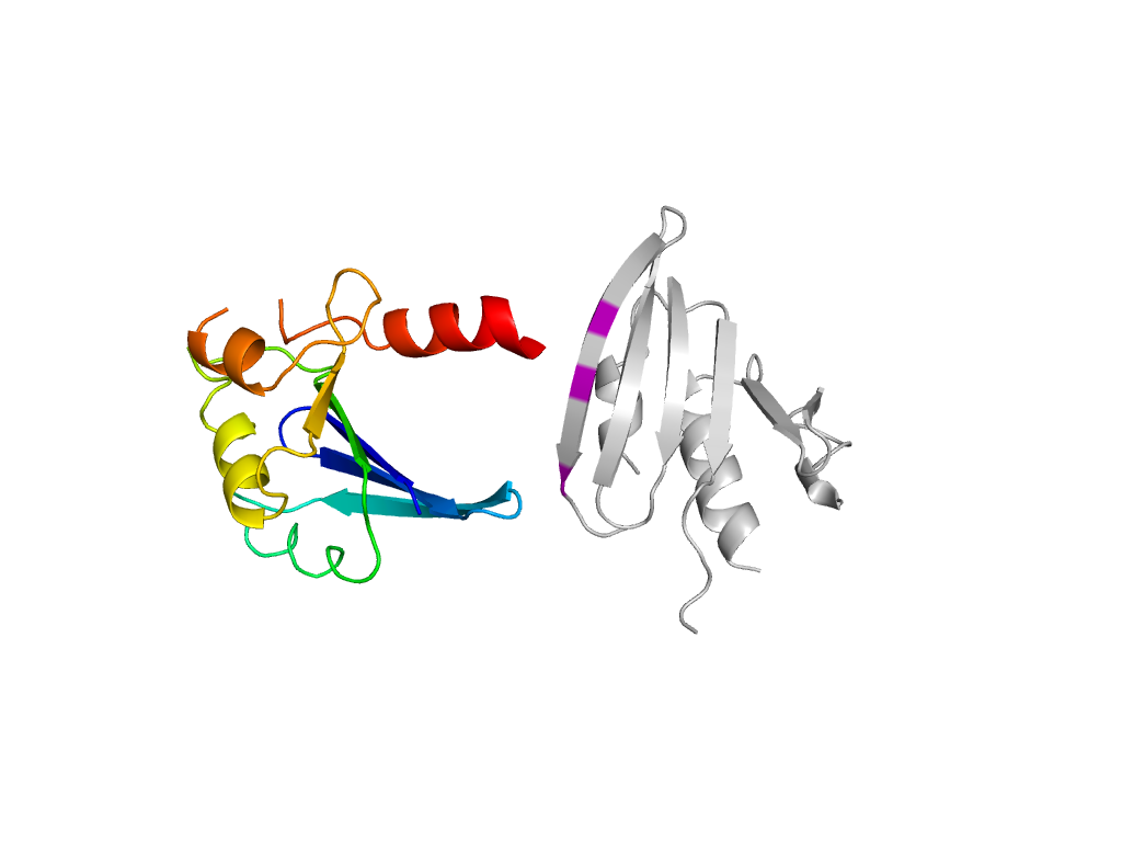
- A: mixed a+b and a/b
- X: Ribonuclease H-like
- H: Ribonuclease H-like
- T: Ribonuclease H-like
- F: Hydantoinase_A
| UID: | 001148160 |
|---|---|
| Type: | Manual and representative domain |
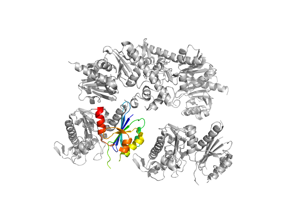
| Range: | A:1-125 |
| PDB: | 3c0b |
| PDB Description: | Conserved archaeal protein Q6M145 |
| UniProt: | Q6M145 |
| Species: |


