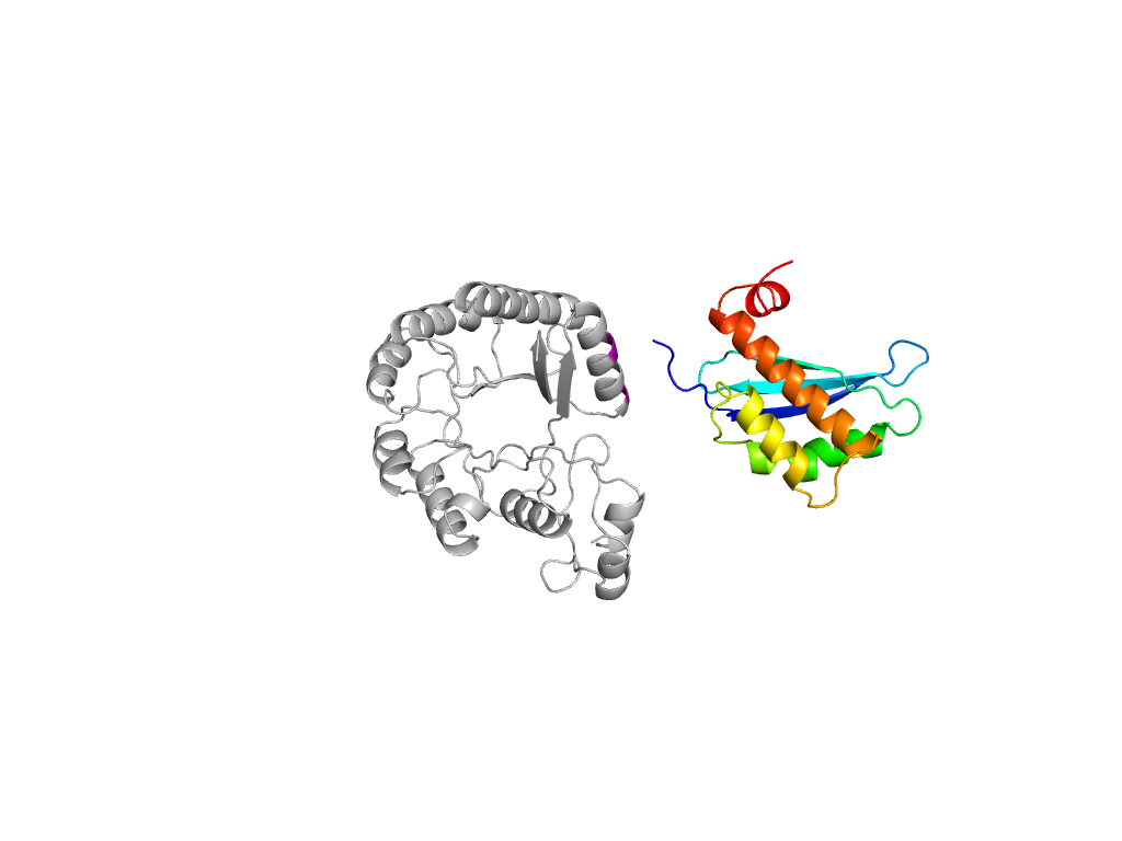
- A: a+b two layers
- X: Enolase-N/ribosomal protein
- H: Enolase N-terminal domain-like
- T: Enolase N-terminal domain-like
- F: MR_MLE_N
| UID: | 000168256 |
|---|---|
| Type: | Automatic domain |
| Parent: | e1mucA1 |
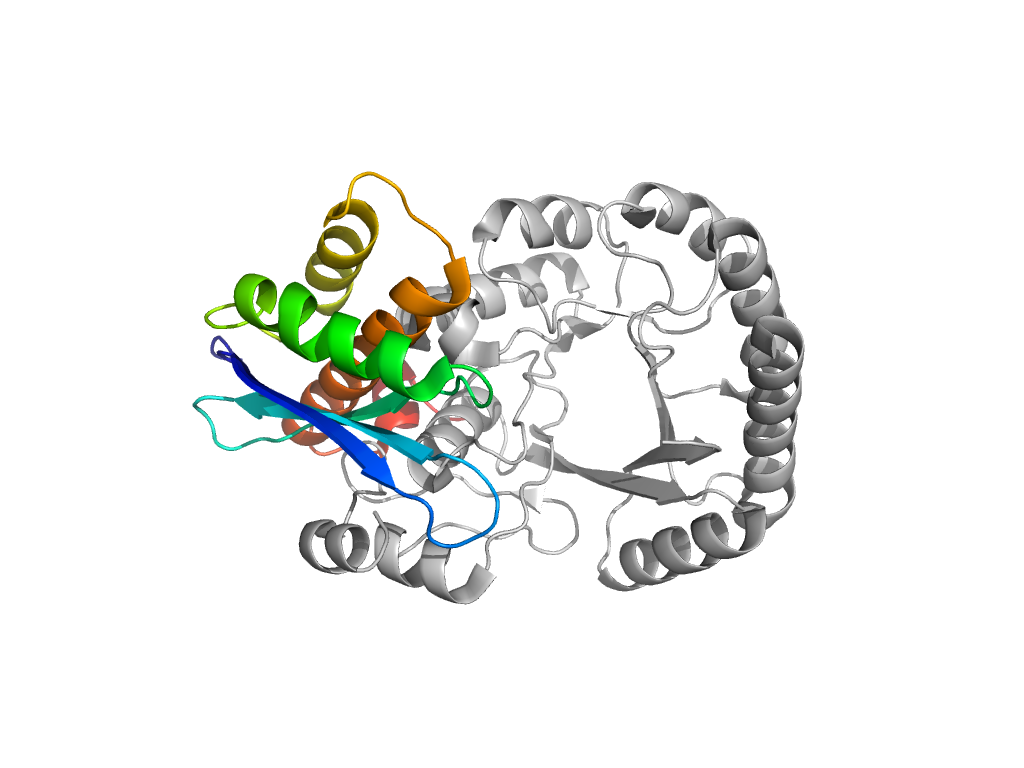
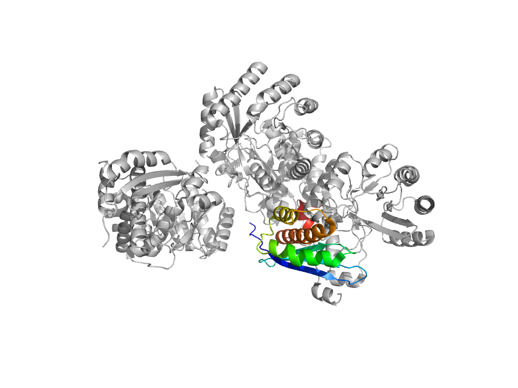
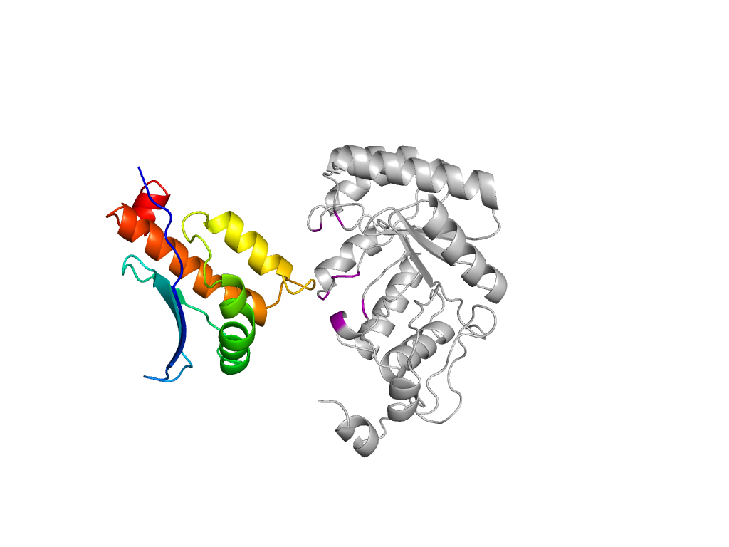
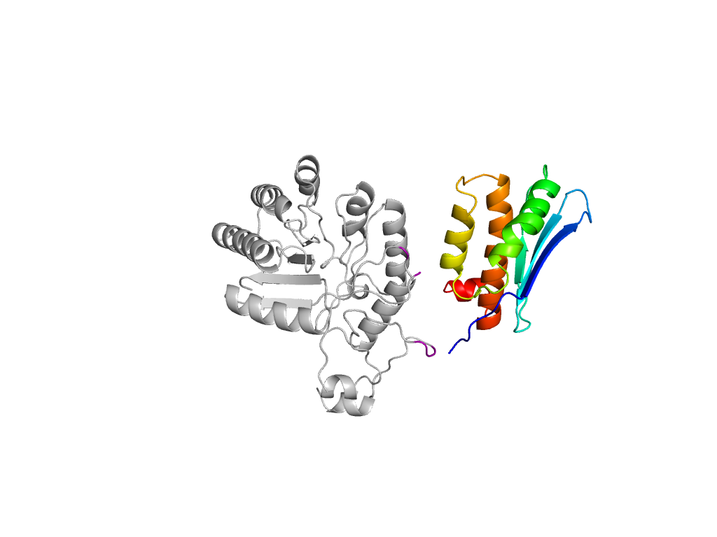
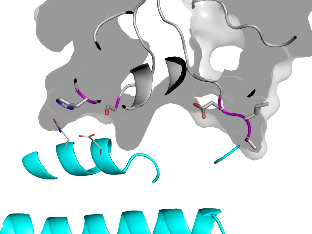
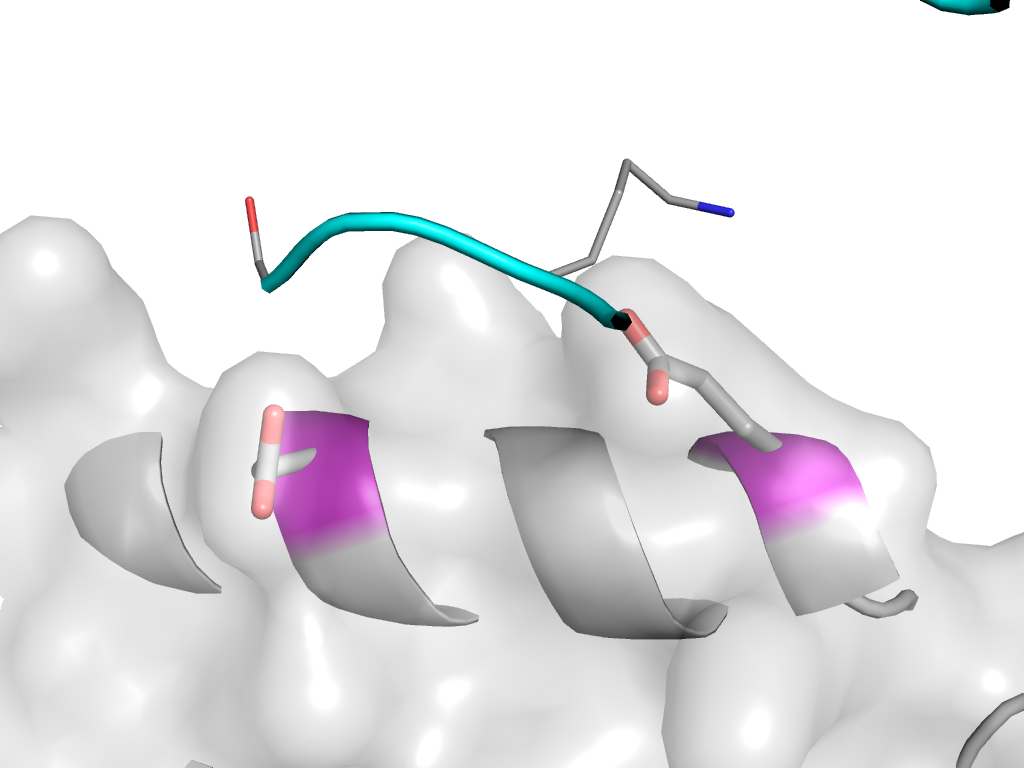
| Range: | A:2-118 |
| PDB: | 3dfh |
| PDB Description: | mandelate racemase |
| UniProt: | A5KUH4 |
| Species: | Vibrionales bacterium SWAT-3 |
