- A: a+b two layers
- X: Alpha-beta plaits
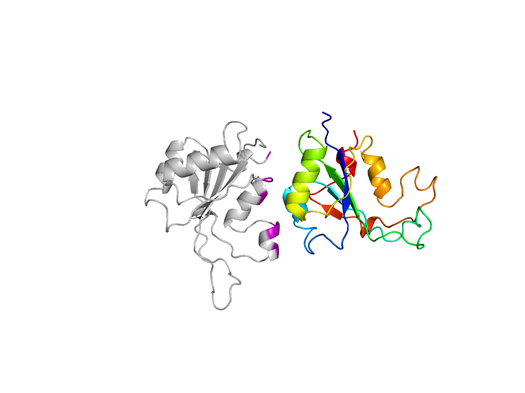
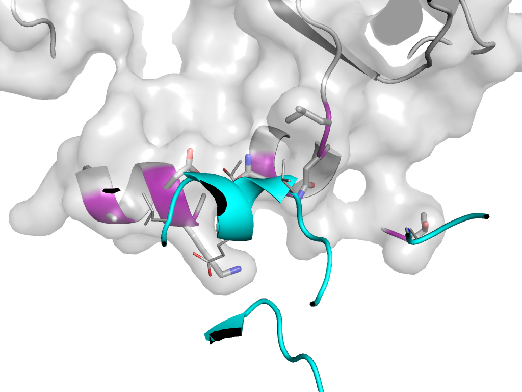
- H: Origin of replication-binding domains
- T: Origin of replication-binding domains
- F: Rep_2
| UID: | 000135397 |
|---|---|
| Type: | Manual and representative domain |
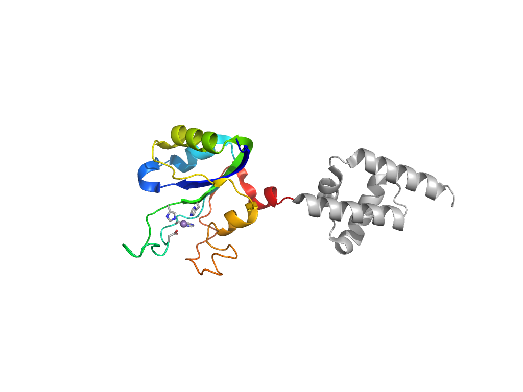
| Range: | A:3-133 |
| Ligands: | MN |
| PDB: | 3dkx |
| PDB Description: | Replication protein repB |
| UniProt: | P13921 |
| Species: | Streptococcus agalactiae |

