- A: a/b three-layered sandwiches
- X: Periplasmic binding protein-like II
- H: Periplasmic binding protein-like II
- T: Periplasmic binding protein-like II
- F: SBP_bac_1
| UID: | 001520163 |
|---|---|
| Type: | Automatic domain |
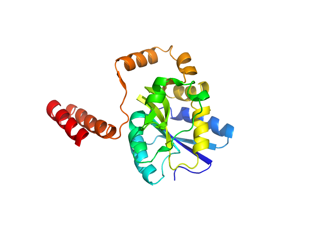
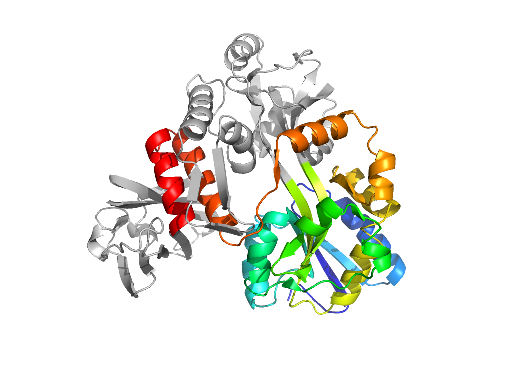
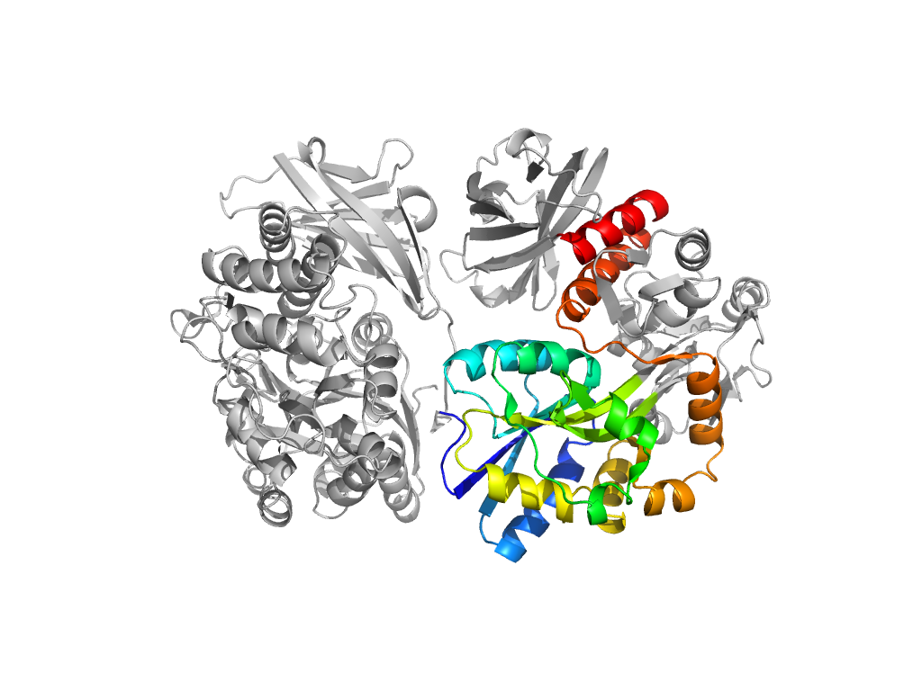
| Parent: | e1laxA2 |
| Range: | A:1-112,A:261-371 |
| Ligands: | CA MAL ZN |
| PDB: | 3ef7 |
| PDB Description: | Maltose-binding periplasmic protein, LINKER, Zona pellucida protein 3 |
| Species: | Mus musculus |
