| Domain ID | Symmetry operator | H-group | Visualization |
|---|
| e3egwB1 |
A:X,Y,Z->B:x,y,z |
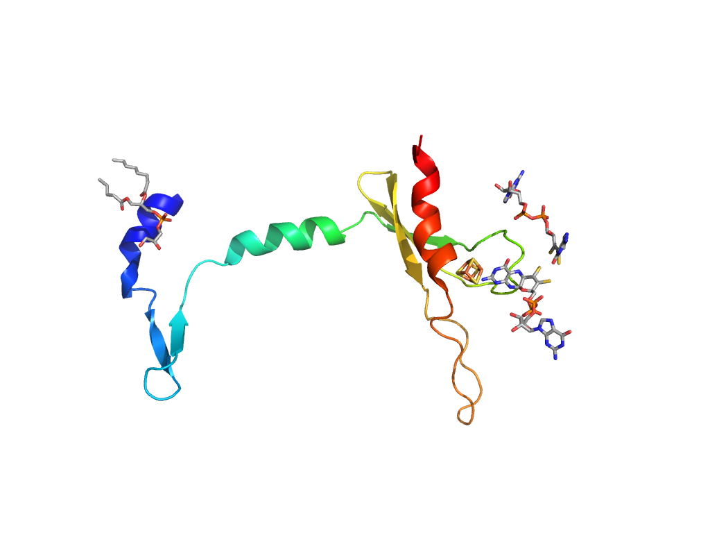
4Fe-4S ferredoxin |
Interaction
Interface
Pymol
|
| e3egwB2 |
A:X,Y,Z->B:x,y,z |
4Fe-4S ferredoxin |
Interaction
Interface
Pymol
|
| e3egwA4 |
A:x,y,z->A:-X-2,Y,-Z-1/2 |
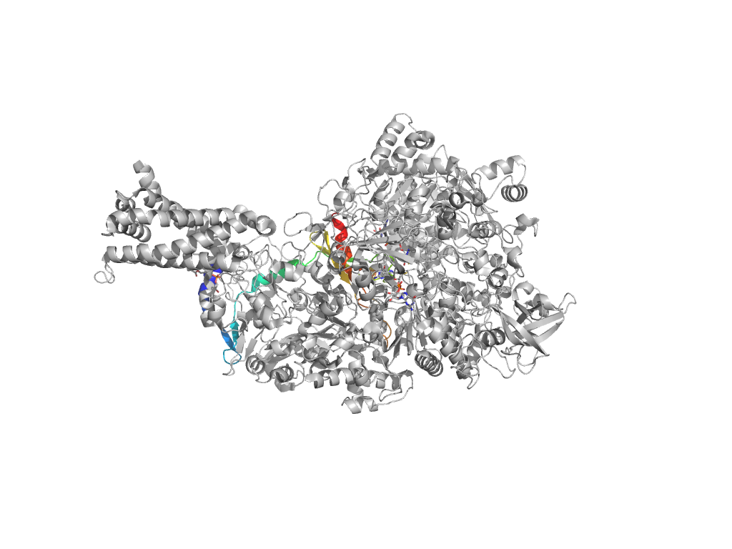
Formate dehydrogenase/DMSO reductase, domain 1 |
Interaction
Interface
Pymol
|
| e3egwA2 |
A:x,y,z->A:-X-2,Y,-Z-1/2 |
Formate dehydrogenase/DMSO reductase, domains 2 and 3 |
Interaction
Interface
Pymol
|
| e3egwA4 |
A:-X-2,Y,-Z-1/2->A:x,y,z |
Formate dehydrogenase/DMSO reductase, domain 1 |
Interaction
Interface
Pymol
|
| e3egwA2 |
A:-X-2,Y,-Z-1/2->A:x,y,z |
Formate dehydrogenase/DMSO reductase, domains 2 and 3 |
Interaction
Interface
Pymol
|
| e3egwB1 |
A:-X-2,Y,-Z-1/2->B:x,y,z |
4Fe-4S ferredoxin |
Interaction
Interface
Pymol
|
| e3egwC1 |
A:X,Y,Z->C:x,y,z |
Transmembrane heme-binding four-helical bundle |
Interaction
Interface
Pymol
|
| e3egwA2 |
A:x,y,z->A:X-1/2,-Y-1/2,-Z |
Formate dehydrogenase/DMSO reductase, domains 2 and 3 |
Interaction
Interface
Pymol
|

