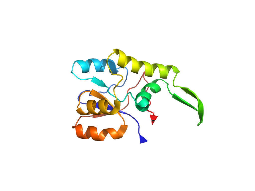
- A: a/b three-layered sandwiches
- X: Periplasmic binding protein-like II
- H: Periplasmic binding protein-like II
- T: Periplasmic binding protein-like II
- F: SBP_bac_1
| UID: | 001654192 |
|---|---|
| Type: | Automatic domain |
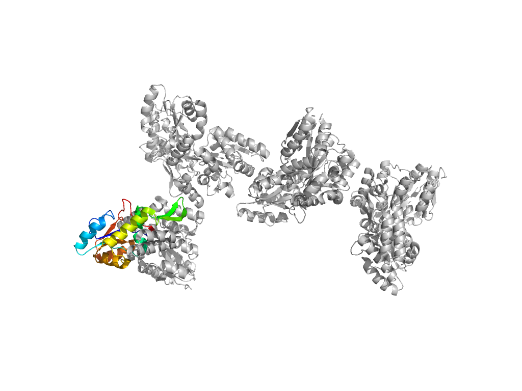
| Parent: | e3waiA4 |
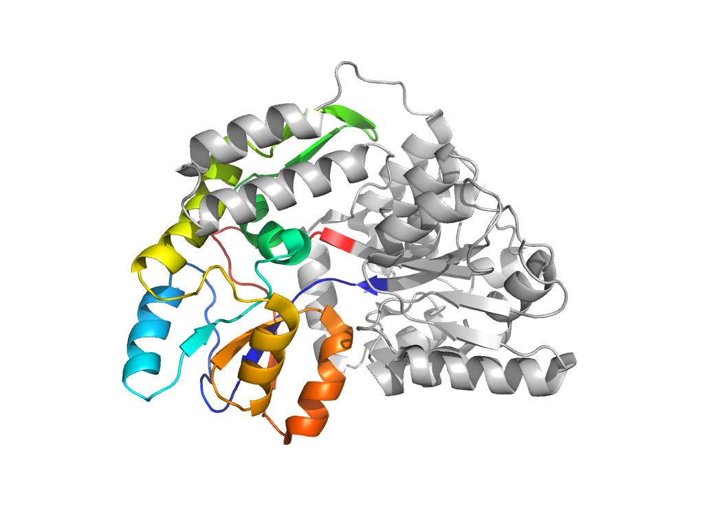
| Range: | D:110-259 |
| Ligands: | MAL SO4 |
| PDB: | 3g7v |
| PDB Description: | Maltose-binding periplasmic protein, Islet amyloid polypeptide fusion protein |
| UniProt: | P0AEX9 |
| Species: | Homo sapiens |
