Domain ID e3gvlA1

Download files:
  • A: beta duplicates or obligate multimers
  • X: Single-stranded right-handed beta-helix
  • H: Pectin lyase-like
  • T: Pectin lyase-like
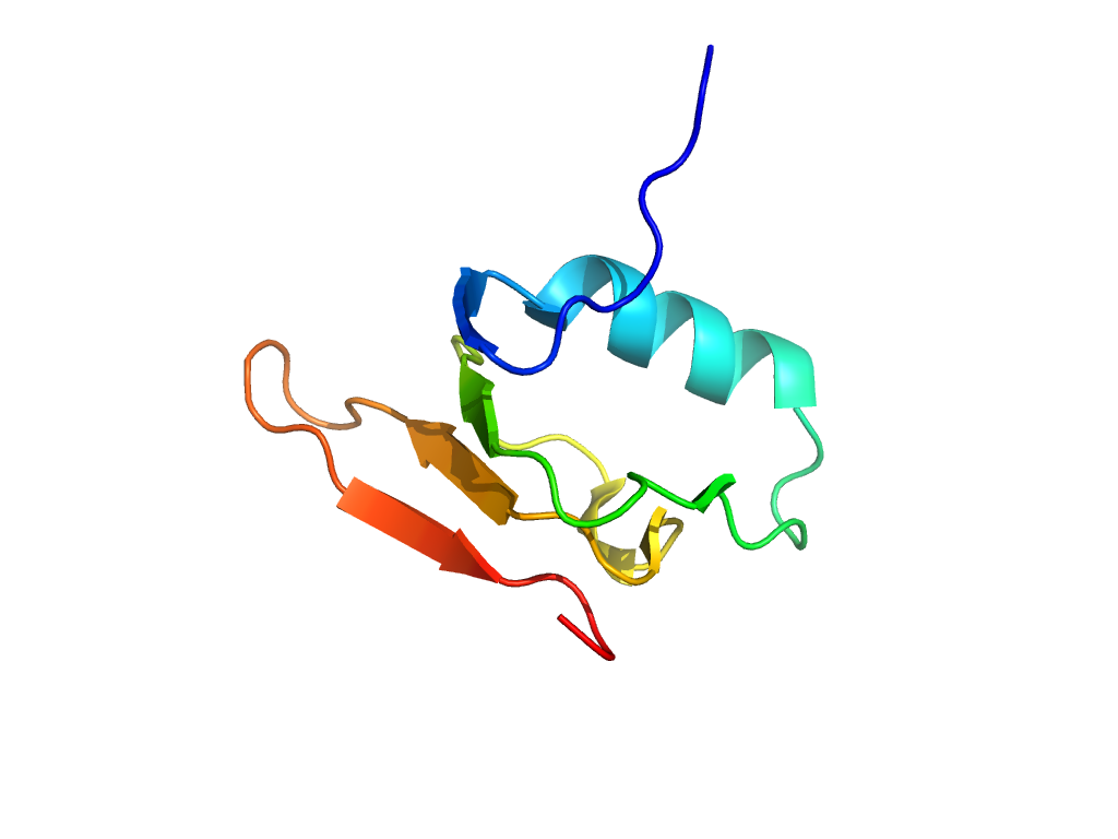
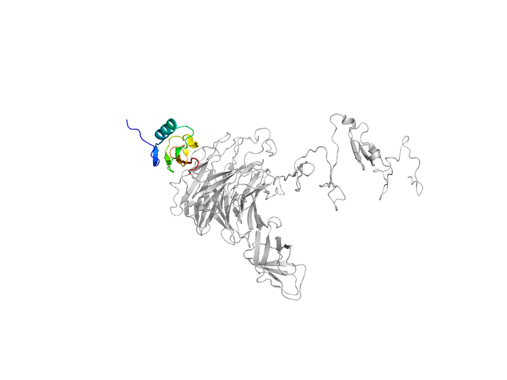
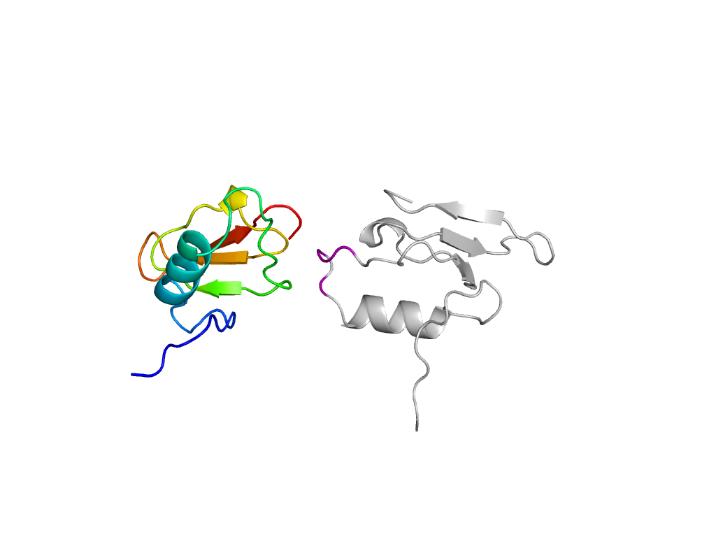
  • F: End_N_terminal
UID: 001338945
Type: Automatic domain
Parent: e1v0eA5
Range: A:241-311
PDB: 3gvl
PDB Description: Endo-N-acetylneuraminidase
UniProt: Q04830
Species: Enterobacteria phage K1F

Structure of domain e3gvlA1

Structural contacts of domain e3gvlA1 of H-group "Pectin lyase-like":

The domain and its neighboring domains (both within the asymmetric unit and to crystallographic symmetry mates) are represented by spheres and linked by lines. Distances between the center of the domain and interfaces are shown for each contact.

Domain IDSymmetry operatorH-groupVisualization
e3gvlA1 A:x,y,z->A:-Y+1,X-Y,Z Pectin lyase-like Interaction Interface Pymol
e3gvlA2 A:-Y+1,X-Y,Z->A:x,y,z Phage tail fiber protein trimerization domain Interaction Interface Pymol
e3gvlA1 A:-Y+1,X-Y,Z->A:x,y,z Pectin lyase-like Interaction Interface Pymol
e3gvlA4 A:-Y+1,X-Y,Z->A:x,y,z beta-propeller Interaction Interface Pymol
e3gvlA3 A:x,y,z->A:-Y+2/3,X-Y+1/3,Z+1/3 RIFT-related Interaction Interface Pymol