Domain ID e3ho7A1

Download files:
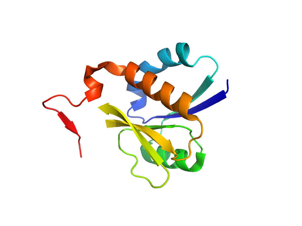
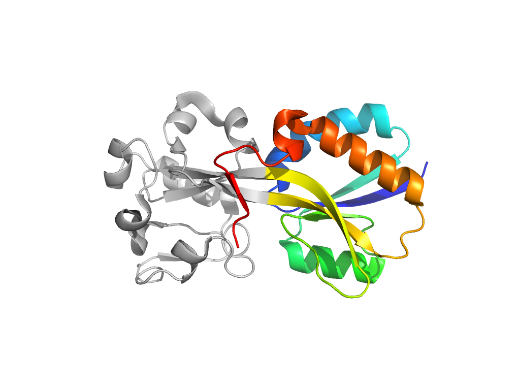
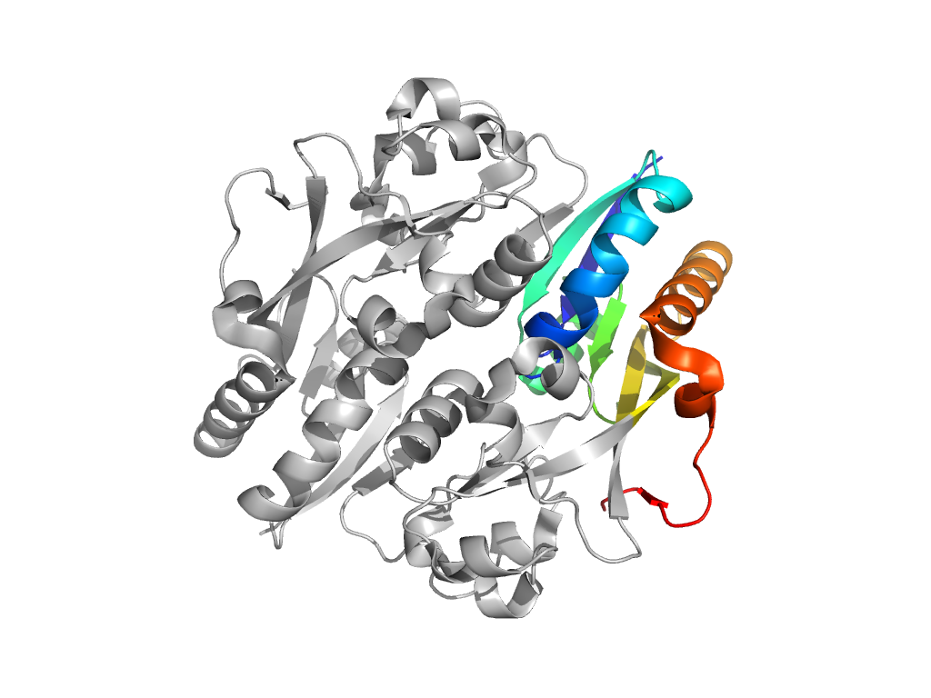
  • A: a/b three-layered sandwiches
  • X: Periplasmic binding protein-like II
  • H: Periplasmic binding protein-like II
  • T: Periplasmic binding protein-like II
  • F: LysR_substrate
UID: 001520842
Type: Automatic domain
Parent: e1i6aA3
Range: A:90-162,A:268-309
PDB: 3ho7
PDB Description: OXYR
UniProt: Q7MXD3
Species: Porphyromonas gingivalis

Structure of domain e3ho7A1

Structural contacts of domain e3ho7A1 of H-group "Periplasmic binding protein-like II":

Sorry, the images are being generated.

The domain and its neighboring domains (both within the asymmetric unit and to crystallographic symmetry mates) are represented by spheres and linked by lines. Distances between the center of the domain and interfaces are shown for each contact.

Domain IDSymmetry operatorH-groupVisualization
e3ho7B1 A:X,Y,Z->B:x,y,z Periplasmic binding protein-like II Interaction Interface Pymol
e3ho7B1 A:x,y,z->B:X,Y-1,Z Periplasmic binding protein-like II Interaction Interface Pymol
e3ho7A2 A:x,y,z->A:X-1,Y-1,Z-1 Periplasmic binding protein-like II Interaction Interface Pymol
e3ho7B2 A:x,y,z->B:X,Y-1,Z-1 Periplasmic binding protein-like II Interaction Interface Pymol