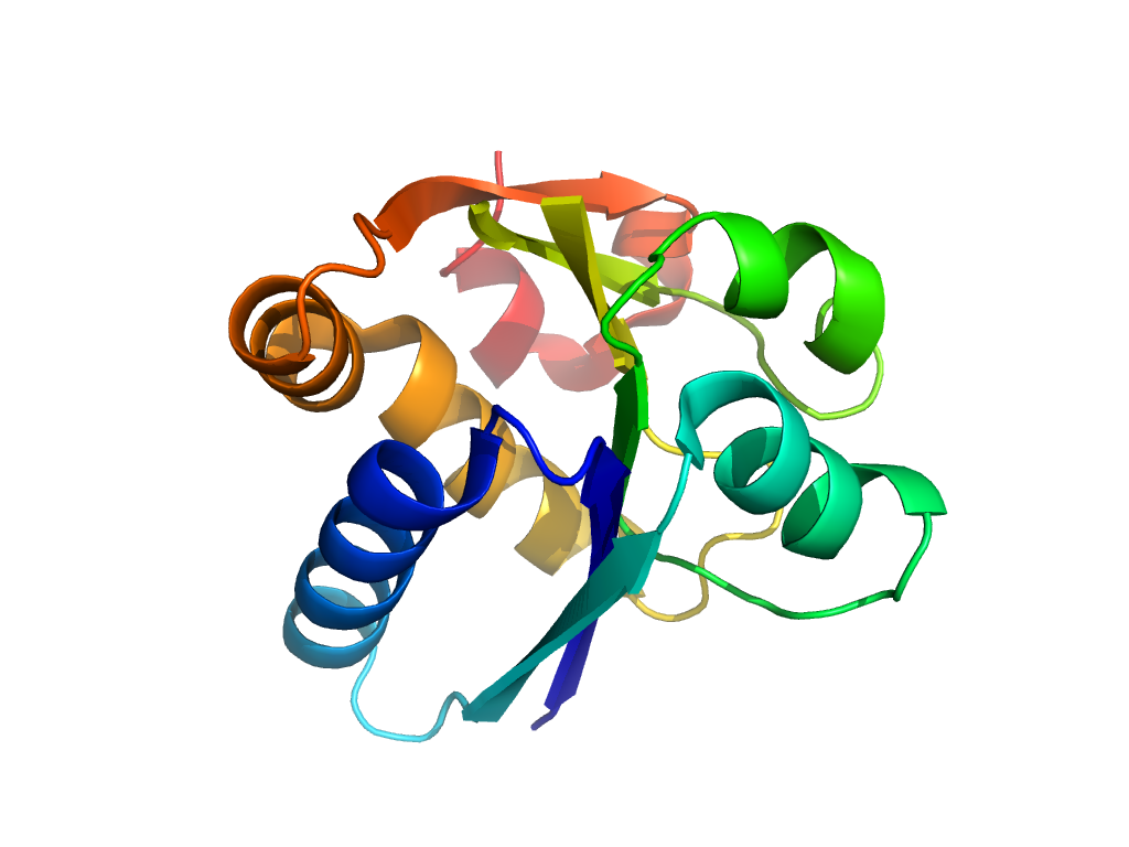
- A: a/b three-layered sandwiches
- X: Periplasmic binding protein-like II
- H: Periplasmic binding protein-like II
- T: Periplasmic binding protein-like II
- F: SBP_bac_11
| UID: | 001521265 |
|---|---|
| Type: | Automatic domain |
| Parent: | e2onsA3 |
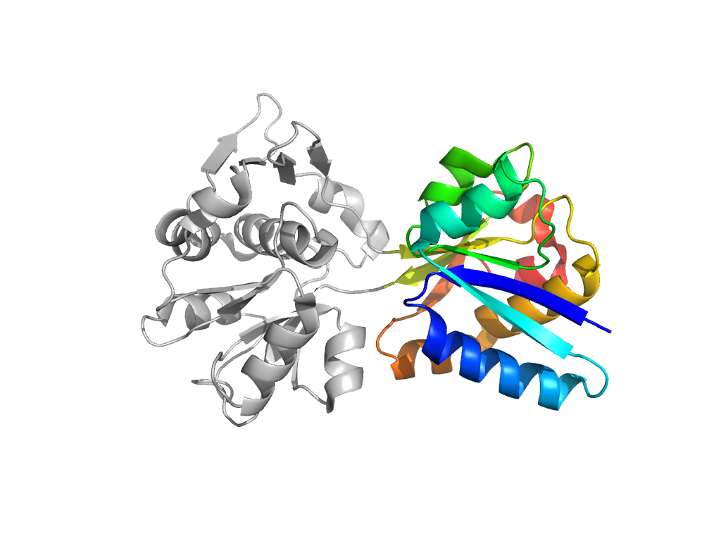
| Range: | A:41-120,A:293-352 |
| Ligands: | CIT |
| PDB: | 3k6v |
| PDB Description: | Solute-binding protein MA_0280 |
| UniProt: | Q8TTZ5 |
| Species: | Methanosarcina acetivorans |

