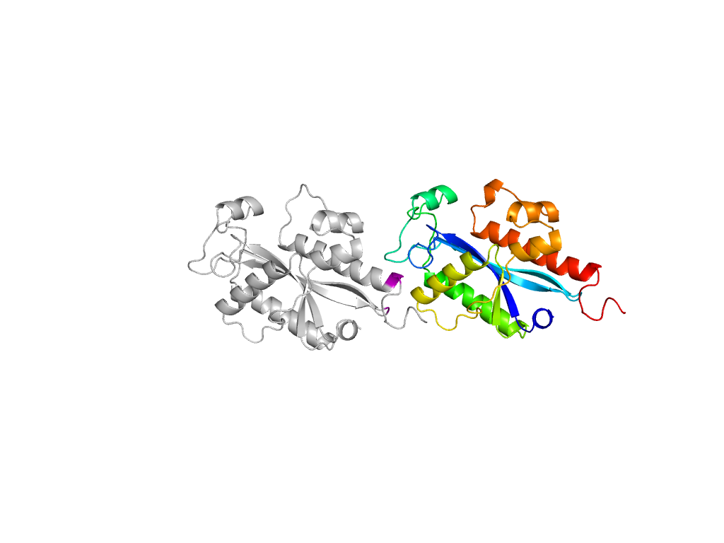
- A: mixed a+b and a/b
- X: Ribonuclease H-like
- H: Ribonuclease H-like
- T: Ribonuclease H-like
- F: RNase_T
| UID: | 000412831 |
|---|---|
| Type: | Automatic domain |
| Parent: | e2igiA1 |
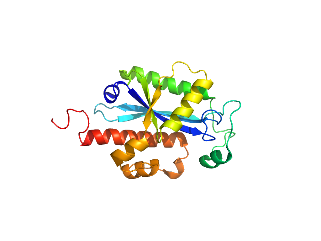
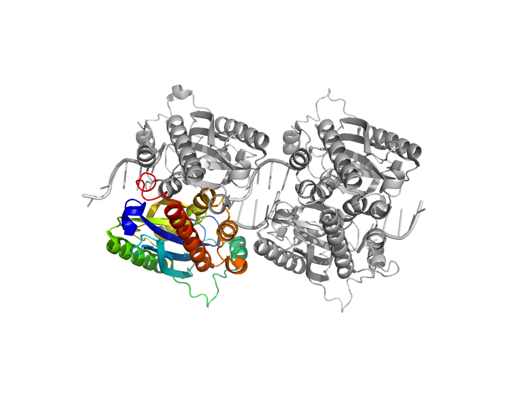
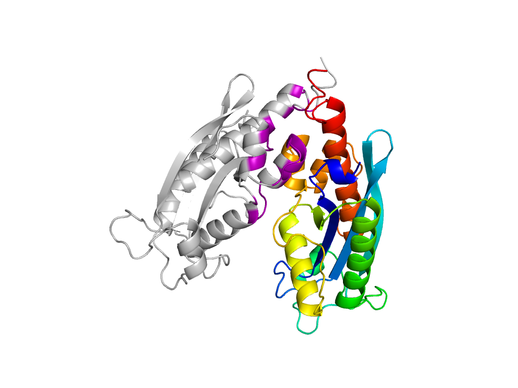
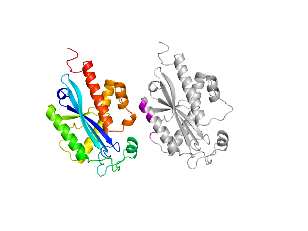
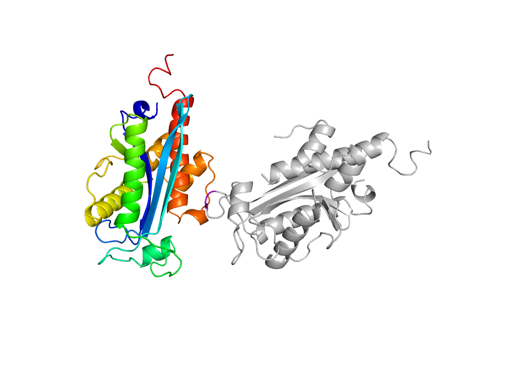
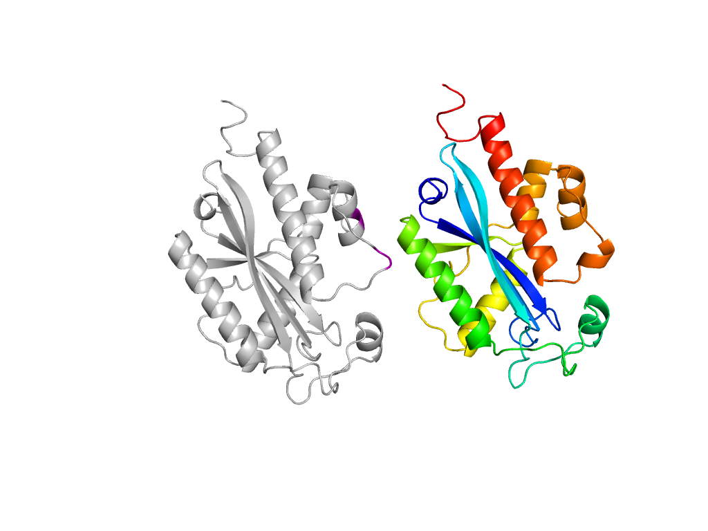
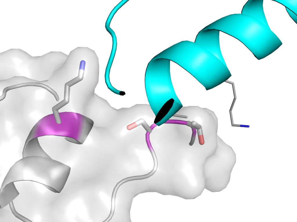
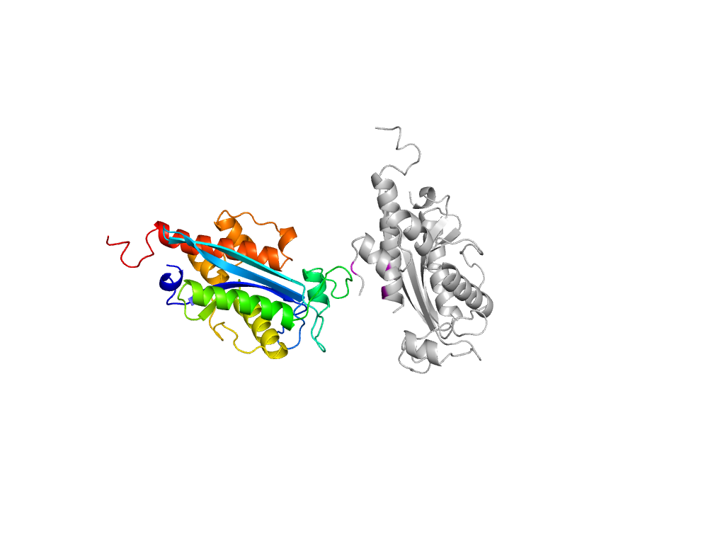
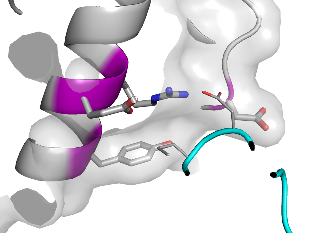
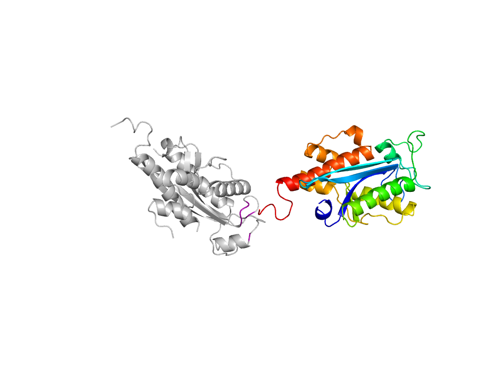
| Range: | E:9-214 |
| PDB: | 3nh2 |
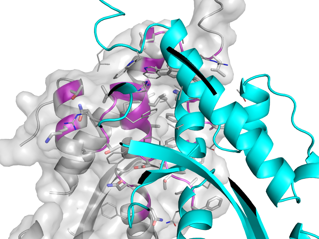
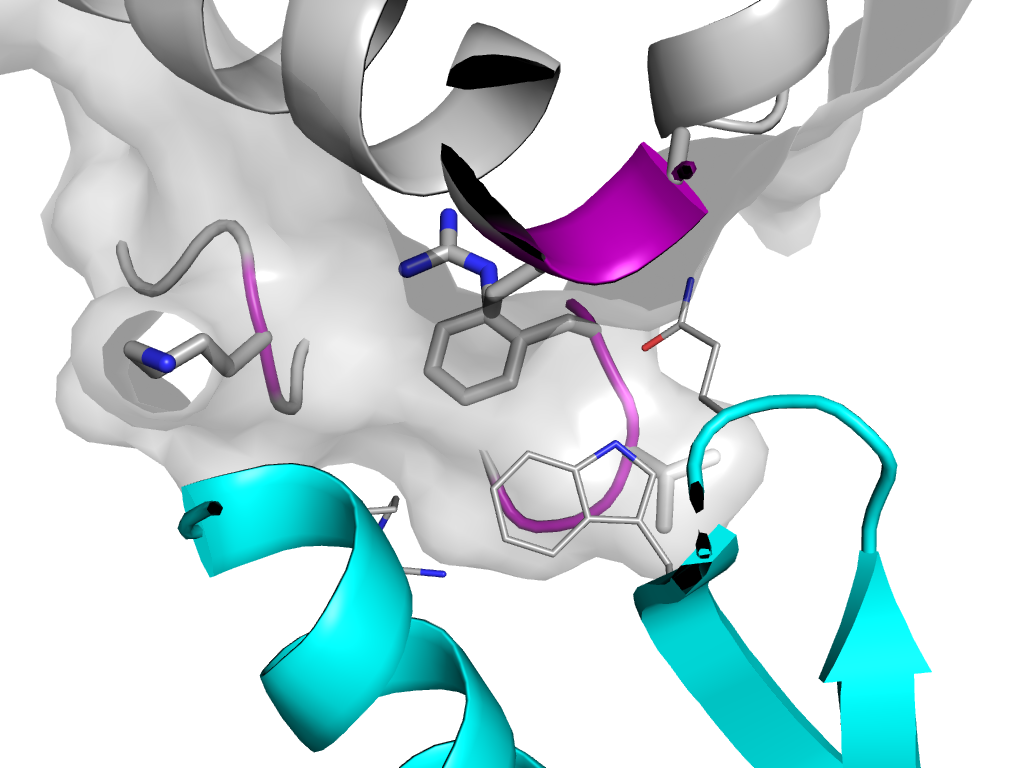
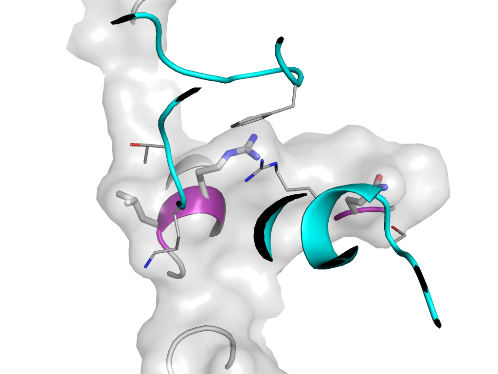
| PDB Description: | RIBONUCLEASE T |
| UniProt: | P30014 |
| Species: | Escherichia coli |
