| Domain ID | Symmetry operator | H-group | Visualization |
|---|
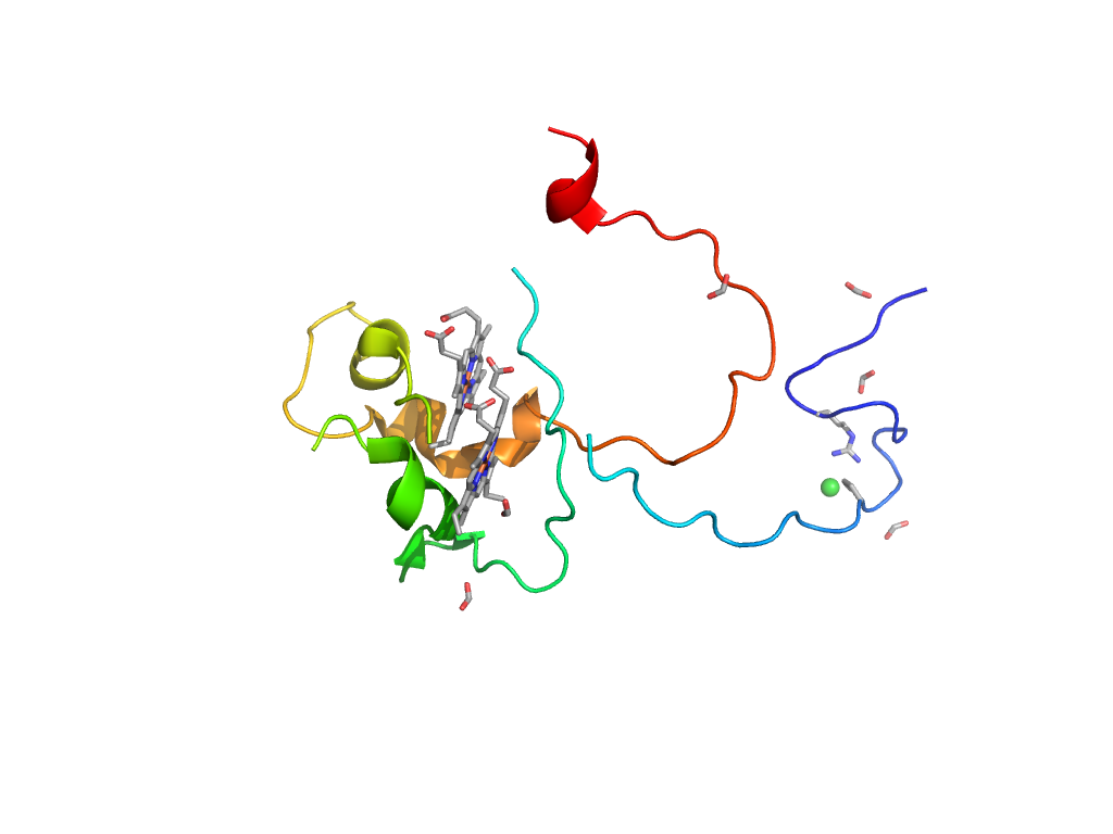
| e3o5aA1 |
B:x,y,z->A:X,Y,Z |
RIFT-related |
Interaction
Interface
Pymol
|
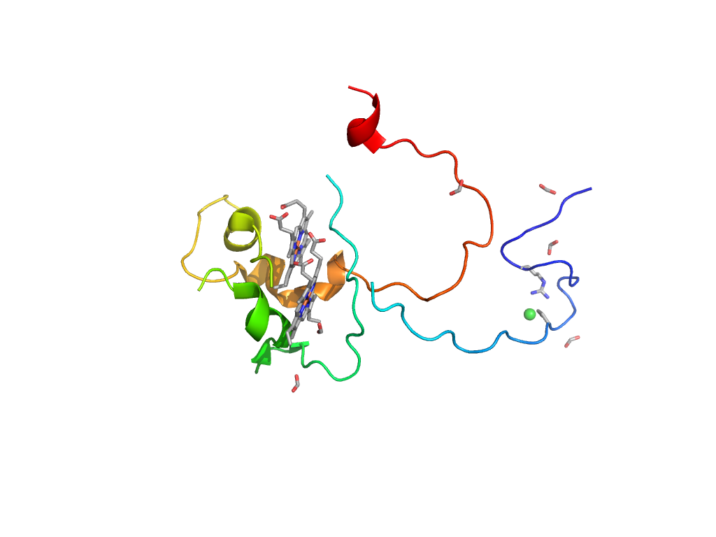
| e3o5aA3 |
B:x,y,z->A:X,Y,Z |
Formate dehydrogenase/DMSO reductase, domains 2 and 3 |
Interaction
Interface
Pymol
|
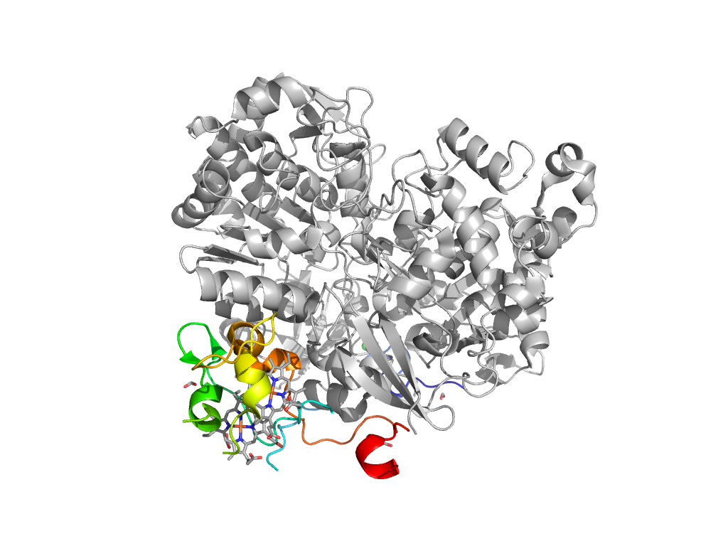
| e3o5aA4 |
B:x,y,z->A:X,Y,Z |
Formate dehydrogenase/DMSO reductase, domain 1 |
Interaction
Interface
Pymol
|
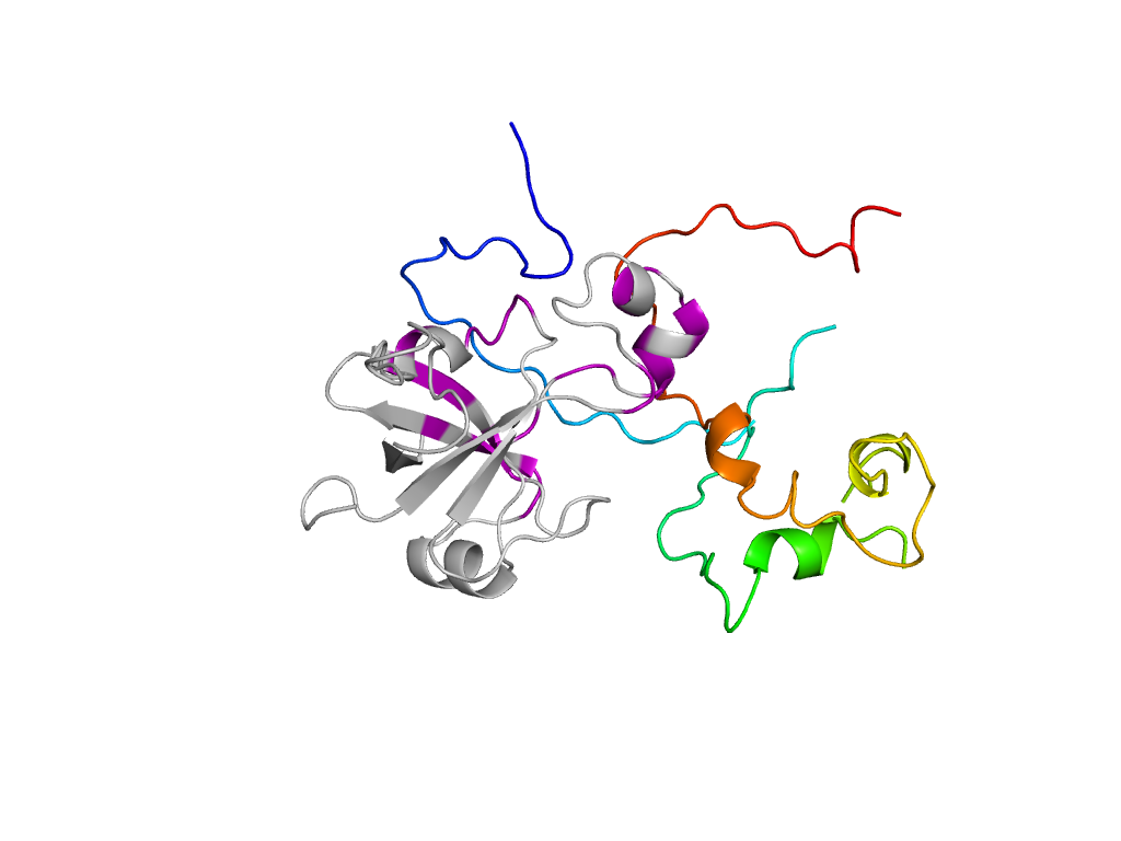
| e3o5aA2 |
B:x,y,z->A:X,Y,Z |
Formate dehydrogenase/DMSO reductase, domains 2 and 3 |
Interaction
Interface
Pymol
|
| e3o5aB1 |
B:x,y,z->B:-X+1,Y,-Z+1 |
Di-heme elbow motif |
Interaction
Interface
Pymol
|
| e3o5aB1 |
B:-X+1,Y,-Z+1->B:x,y,z |
Di-heme elbow motif |
Interaction
Interface
Pymol
|
| e3o5aA4 |
B:x,y,z->A:-X+1,Y,-Z+1 |
Formate dehydrogenase/DMSO reductase, domain 1 |
Interaction
Interface
Pymol
|
| e3o5aA3 |
B:x,y,z->A:-X+1/2,Y-1/2,-Z |
Formate dehydrogenase/DMSO reductase, domains 2 and 3 |
Interaction
Interface
Pymol
|
| e3o5aA2 |
B:X-1/2,Y+1/2,Z->A:x,y,z |
Formate dehydrogenase/DMSO reductase, domains 2 and 3 |
Interaction
Interface
Pymol
|
