| UID: | 001117667 |
|---|---|
| Type: | Manual and representative domain |
| Range: | A:196-445 |
| PDB: | 3pnt |
| PDB Description: | NAD+-GLYCOHYDROLASE |
| UniProt: | D7S065 |
| Species: | Streptococcus pyogenes |


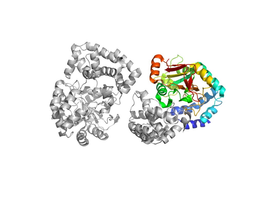
Structure of domain e3pntA1
Domains in the same chain:
| e3pntA1 | A:196-445 | ADP-ribosylation | ADP-ribosylation | ADP-ribosylation |
Domains in the same PDB:
Structural contacts of domain e3pntA1 of H-group "ADP-ribosylation":

The domain and its neighboring domains (both within the asymmetric unit and to crystallographic symmetry mates) are represented by spheres and linked by lines. Distances between the center of the domain and interfaces are shown for each contact.
| Domain ID | Symmetry operator | H-group | Visualization |
|---|---|---|---|
| e3pntB1 | A:X,Y,Z->B:x,y,z | Immunity Factor for SPN | Interaction Interface Pymol |
| e3pntD1 | A:-X+1,Y,-Z+1->D:x,y,z | Immunity Factor for SPN | Interaction Interface Pymol |
| e3pntB1 | A:X,Y-1,Z->B:x,y,z | Immunity Factor for SPN | Interaction Interface Pymol |
| e3pntA1 | A:x,y,z->A:-X+1,Y,-Z+2 | ADP-ribosylation | Interaction Interface Pymol |
| e3pntA1 | A:-X+1,Y,-Z+2->A:x,y,z | ADP-ribosylation | Interaction Interface Pymol |
| e3pntD1 | A:X,Y,Z->D:x,y,z | Immunity Factor for SPN | Interaction Interface Pymol |
| e3pntB1 | A:-X+1,Y,-Z+2->B:x,y,z | Immunity Factor for SPN | Interaction Interface Pymol |
| e3pntC1 | A:-X+1,Y,-Z+1->C:x,y,z | ADP-ribosylation | Interaction Interface Pymol |
| e3pntC1 | A:-X+1,Y-1,-Z+1->C:x,y,z | ADP-ribosylation | Interaction Interface Pymol |