Domain ID e3puvE1

Download files:
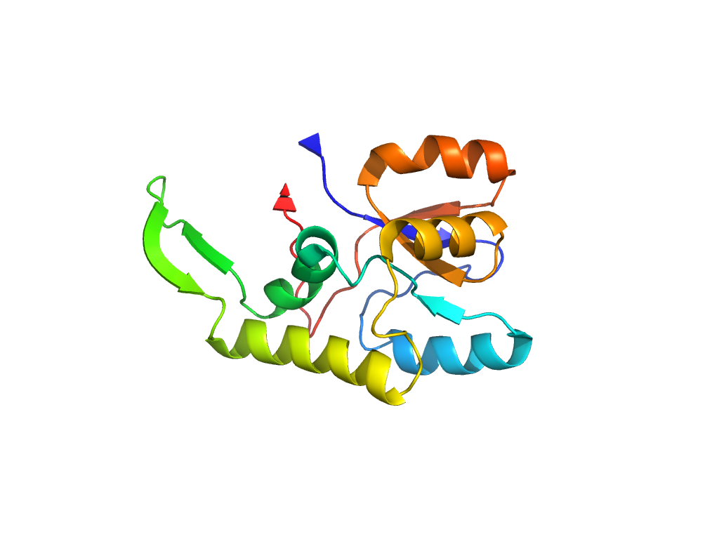
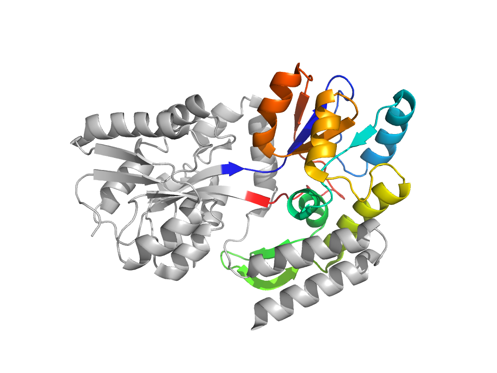
  • A: a/b three-layered sandwiches
  • X: Periplasmic binding protein-like II
  • H: Periplasmic binding protein-like II
  • T: Periplasmic binding protein-like II
  • F: SBP_bac_1
UID: 001522190
Type: Automatic domain
Parent: e4pqkB3
Range: E:110-259
PDB: 3puv
PDB Description: MALTOSE-BINDING PERIPLASMIC PROTEIN
UniProt: P0AEX9
Species:

Structure of domain e3puvE1

Domains in the same chain:

e3puvE2 E:1-109,E:260-370 Periplasmic binding protein-like II Periplasmic binding protein-like II Periplasmic binding protein-like II
e3puvE1 E:110-259 Periplasmic binding protein-like II Periplasmic binding protein-like II Periplasmic binding protein-like II

Domains in the same PDB:

e3puvA2 A:2-235 P-loop domains-like P-loop domains-related P-loop containing nucleoside triphosphate hydrolases
e3puvA4 A:236-372 OB-fold MOP-like MOP-like
e3puvB1 B:2-235 P-loop domains-like P-loop domains-related P-loop containing nucleoside triphosphate hydrolases
e3puvB4 B:236-369 OB-fold MOP-like MOP-like
e3puvE2 E:1-109,E:260-370 Periplasmic binding protein-like II Periplasmic binding protein-like II Periplasmic binding protein-like II
e3puvE1 E:110-259 Periplasmic binding protein-like II Periplasmic binding protein-like II Periplasmic binding protein-like II
e3puvF2 F:10-61 Transmembrane helices in MalF N-terminal region Transmembrane helices in MalF N-terminal region Transmembrane helices in MalF N-terminal region
e3puvF1 F:62-93,F:254-503 Type II ABC exporter transmembrane domain fold Type I ABC importer transmembrane domain fold Type I ABC importer transmembrane domain fold
e3puvF3 F:93-115,F:207-260 linker domain in MalF N-terminal region linker domain in MalF N-terminal region linker domain in MalF N-terminal region
e3puvF4 F:116-206 barrel domain in MalF N-terminal region barrel domain in MalF N-terminal region barrel domain in MalF N-terminal region
e3puvG1 G:9-296 Type II ABC exporter transmembrane domain fold Type I ABC importer transmembrane domain fold Type I ABC importer transmembrane domain fold

Structural contacts of domain e3puvE1 of H-group "Periplasmic binding protein-like II":

Sorry, the images are being generated.

The domain and its neighboring domains (both within the asymmetric unit and to crystallographic symmetry mates) are represented by spheres and linked by lines. Distances between the center of the domain and interfaces are shown for each contact.

Domain IDSymmetry operatorH-groupVisualization
e3puvA2 E:x,y,z->A:X-1,Y+1,Z P-loop domains-related Interaction Interface Pymol