Domain ID e3py7A4

Download files:
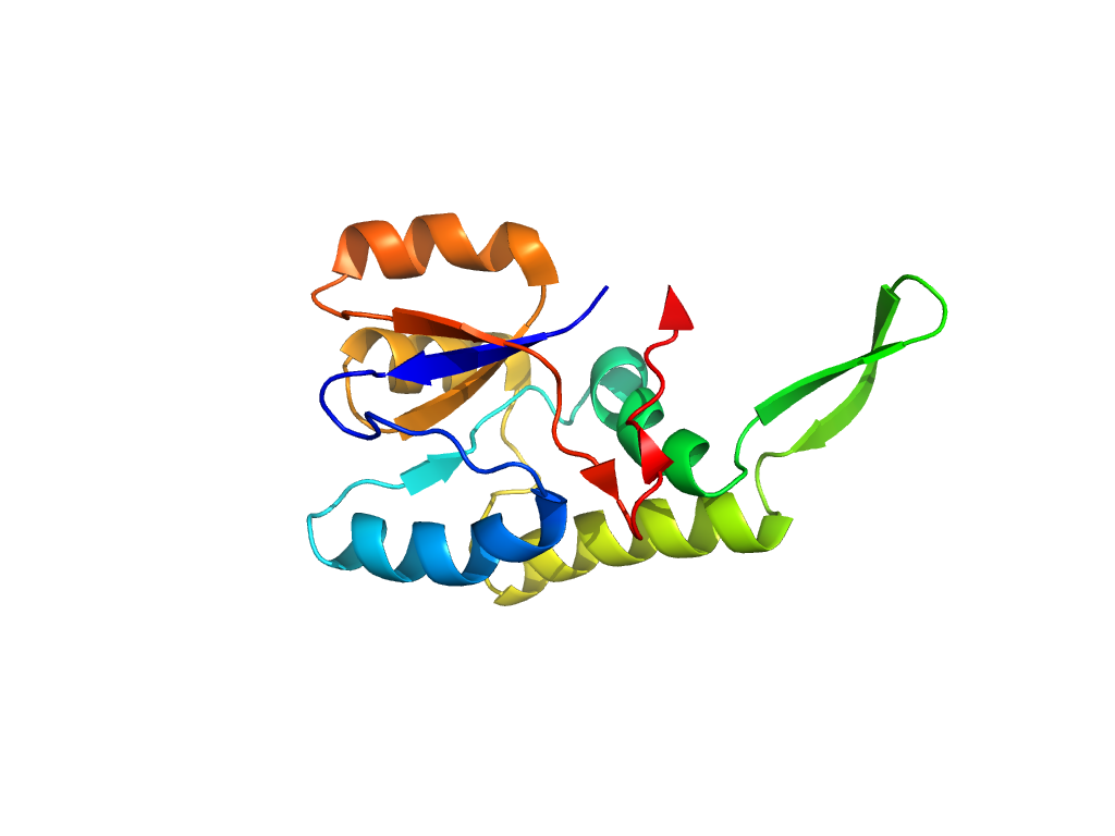
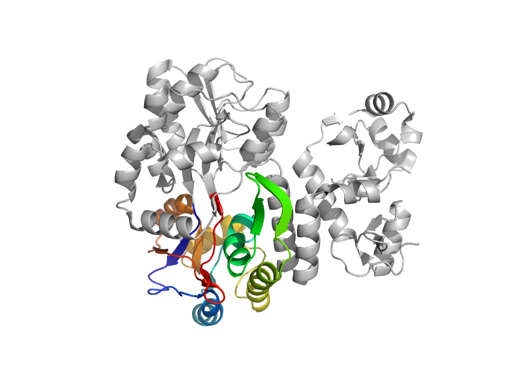
  • A: a/b three-layered sandwiches
  • X: Periplasmic binding protein-like II
  • H: Periplasmic binding protein-like II
  • T: Periplasmic binding protein-like II
  • F: SBP_bac_1
UID: 001518839
Type: Manual and representative domain
Range: A:113-260
Ligands: MLR
PDB: 3py7
PDB Description: maltose-binding periplasmic protein,paxillin LD1,protein E6 chimera
UniProt: P0AEX9
Species: Homo sapiens

Structure of domain e3py7A4

Structural contacts of domain e3py7A4 of H-group "Periplasmic binding protein-like II":

Sorry, the images are being generated.

The domain and its neighboring domains (both within the asymmetric unit and to crystallographic symmetry mates) are represented by spheres and linked by lines. Distances between the center of the domain and interfaces are shown for each contact.

Domain IDSymmetry operatorH-groupVisualization
e3py7A3 A:x,y,z->A:-X+1,Y,-Z+1/2 E6 C-terminal domain-like Interaction Interface Pymol
e3py7A3 A:-X+1,Y,-Z+1/2->A:x,y,z E6 C-terminal domain-like Interaction Interface Pymol
e3py7A4 A:x,y,z->A:X-1/2,-Y+1/2,-Z Periplasmic binding protein-like II Interaction Interface Pymol
e3py7A4 A:X-1/2,-Y+1/2,-Z->A:x,y,z Periplasmic binding protein-like II Interaction Interface Pymol