- A: a+b two layers
- X: Alpha-beta plaits
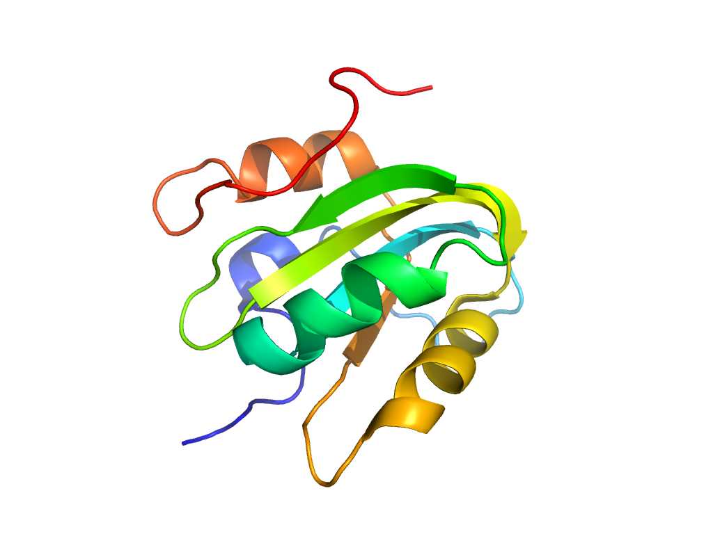
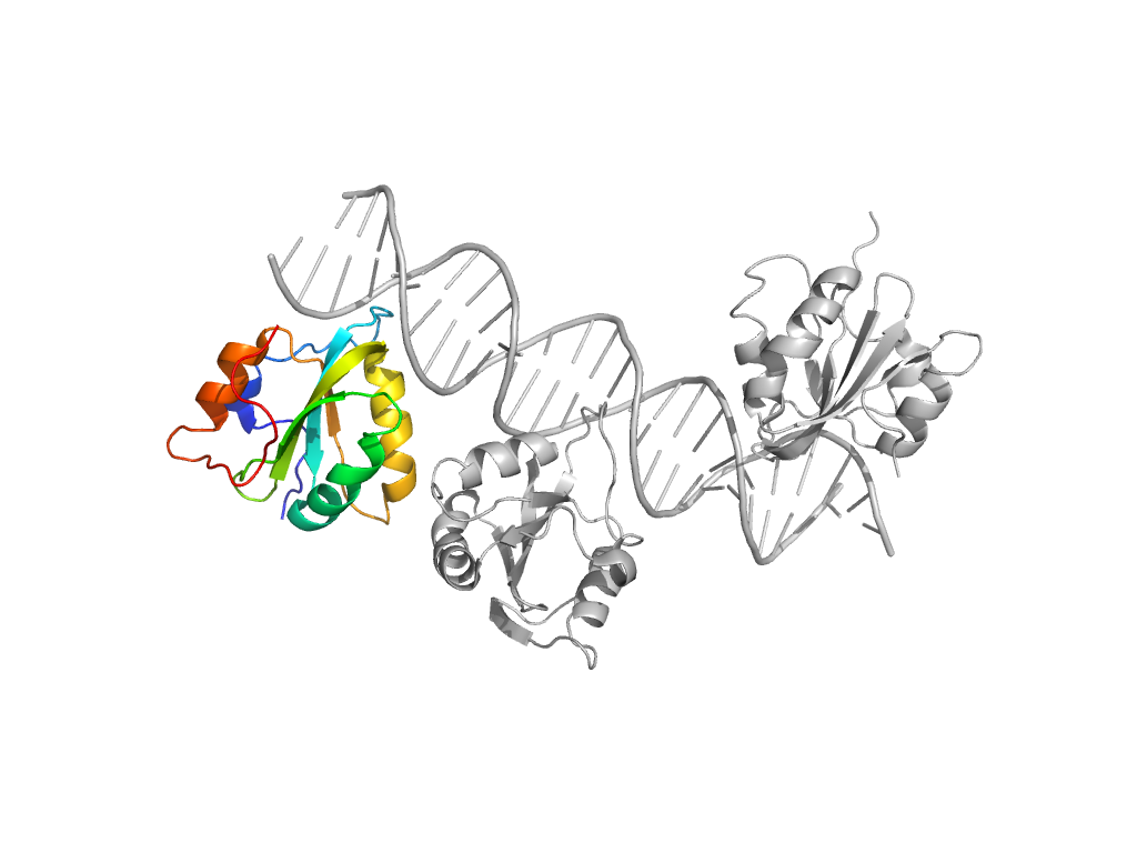
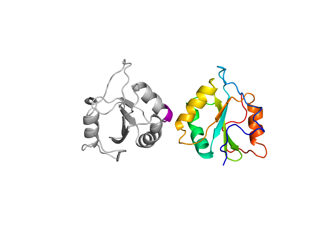
- H: Origin of replication-binding domains
- T: Origin of replication-binding domains
- F: T_Ag_DNA_bind
| UID: | 000418018 |
|---|---|
| Type: | Automatic domain |
| Parent: | e2fufA1 |
| Range: | B:309-428 |
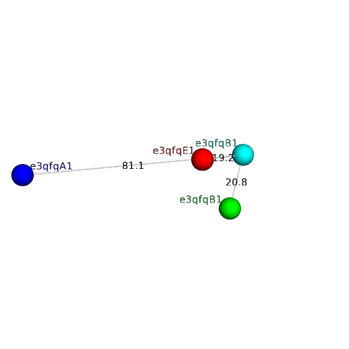
| PDB: | 3qfq |
| PDB Description: | LARGE T ANTIGEN |
| UniProt: | E2IPT4 |
| Species: | Merkel cell polyomavirus |
