| Domain ID | Symmetry operator | H-group | Visualization |
|---|
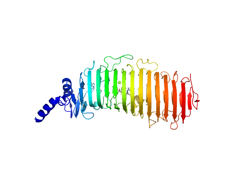
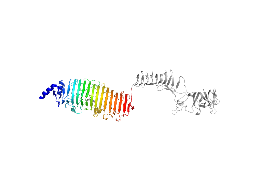
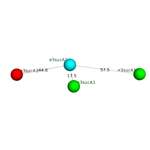
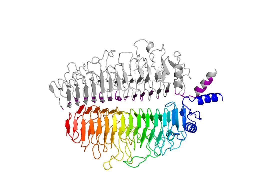
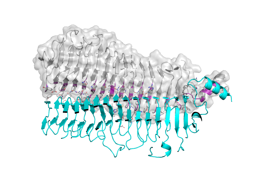
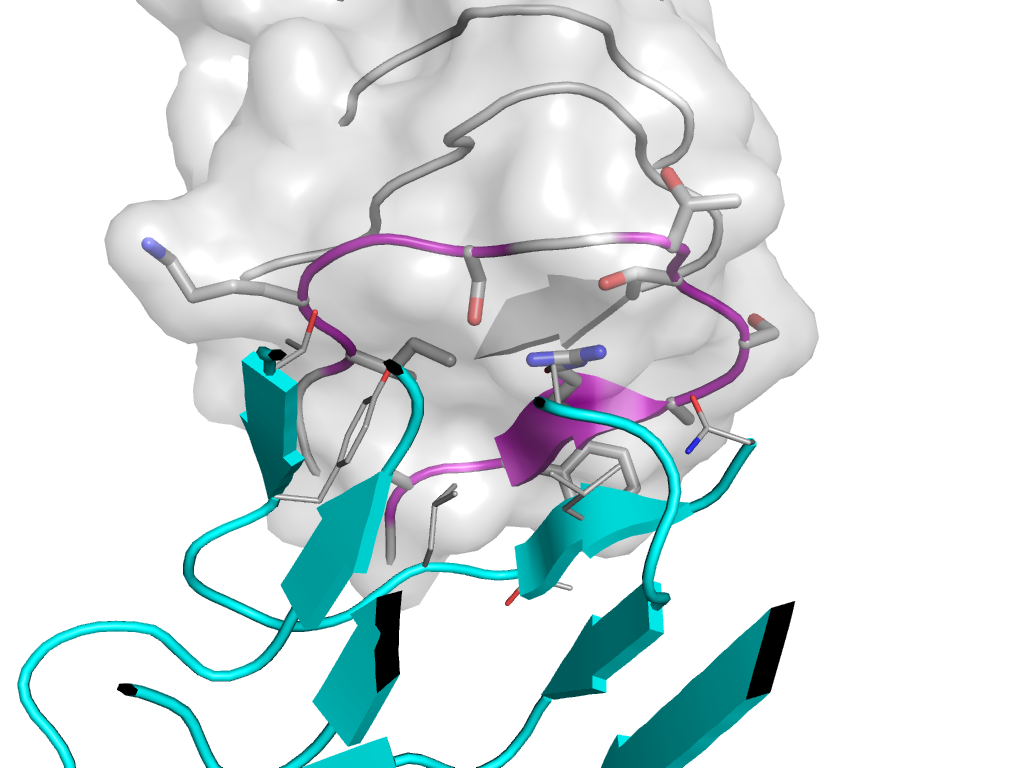
| e3sucA1 |
A:x,y,z->A:-Y,X-Y-1,Z |
Pectin lyase-like |
Interaction
Interface
Pymol
|
| e3sucA3 |
A:x,y,z->A:-Y,X-Y-1,Z |
Adhesin YadA, collagen-binding domain |
Interaction
Interface
Pymol
|
| e3sucA1 |
A:-Y,X-Y-1,Z->A:x,y,z |
Pectin lyase-like |
Interaction
Interface
Pymol
|
| e3sucA1 |
A:x,y,z->A:X-Y-1/3,-Y-2/3,-Z+1/3 |
Pectin lyase-like |
Interaction
Interface
Pymol
|
| e3sucA1 |
A:X-Y-1/3,-Y-2/3,-Z+1/3->A:x,y,z |
Pectin lyase-like |
Interaction
Interface
Pymol
|
| e3sucA1 |
A:x,y,z->A:Y+2/3,X-2/3,-Z+1/3 |
Pectin lyase-like |
Interaction
Interface
Pymol
|
| e3sucA1 |
A:Y+2/3,X-2/3,-Z+1/3->A:x,y,z |
Pectin lyase-like |
Interaction
Interface
Pymol
|
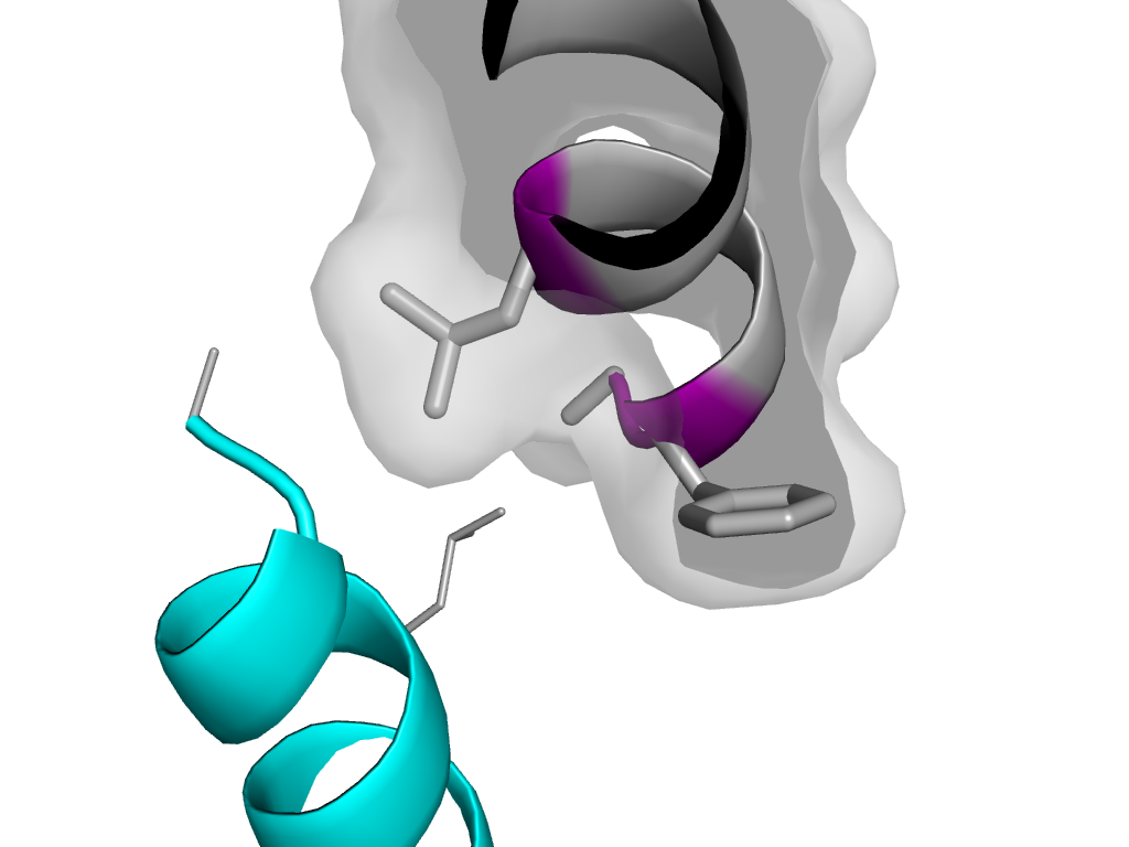
| e3sucA2 |
A:x,y,z->A:X-Y-1,-Y-1,-Z |
Head decoration protein D (gpD, major capsid protein D) |
Interaction
Interface
Pymol
|
