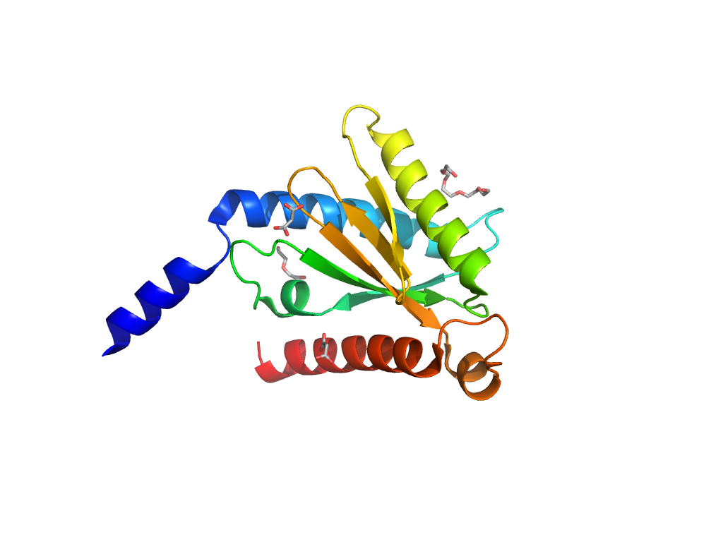
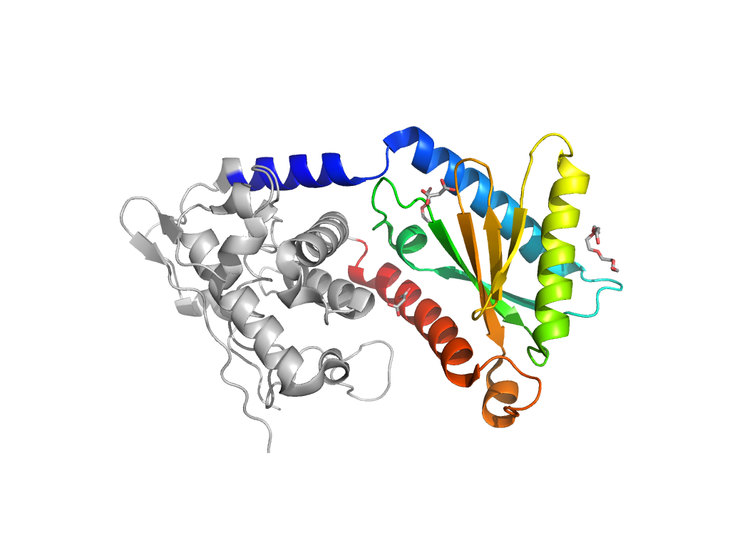
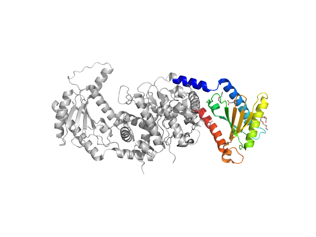
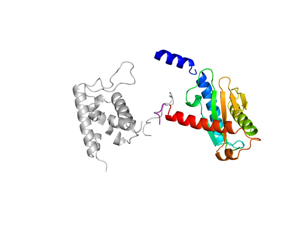
Domain ID e4at7A1

Download files:
  • A: a+b three layers
  • X: Nucleotidyltransferase-like
  • H: Nucleotidyltransferase
  • T: Nucleotidyltransferase
  • F: DZF_N
UID: 001390413
Type: Automatic domain
Parent: e4at9B6
Range: A:53-216
Ligands: 1PE MLI PEG
PDB: 4at7
PDB Description: INTERLEUKIN ENHANCER-BINDING FACTOR 2
UniProt: Q9CXY6
Hsap BLAST neighbor: Q12905
Species: Mus musculus

Structure of domain e4at7A1

Structural contacts of domain e4at7A1 of H-group "Nucleotidyltransferase":

The domain and its neighboring domains (both within the asymmetric unit and to crystallographic symmetry mates) are represented by spheres and linked by lines. Distances between the center of the domain and interfaces are shown for each contact.

Domain IDSymmetry operatorH-groupVisualization
e4at7B1 A:-X+1,Y,-Z->B:x,y,z SAM/DNA-glycosylase Interaction Interface Pymol
e4at7B4 A:-X+1/2,-Y+1/2,Z-1/2->B:x,y,z Nucleotidyltransferase Interaction Interface Pymol
e4at7A2 A:X-1/2,-Y+1/2,-Z+1/2->A:x,y,z SAM/DNA-glycosylase Interaction Interface Pymol
e4at7A1 A:x,y,z->A:-X+1,-Y,Z Nucleotidyltransferase Interaction Interface Pymol
e4at7A1 A:-X+1,-Y,Z->A:x,y,z Nucleotidyltransferase Interaction Interface Pymol