- A: a/b three-layered sandwiches
- X: Periplasmic binding protein-like II
- H: Periplasmic binding protein-like II
- T: Periplasmic binding protein-like II
- F: SBP_bac_1
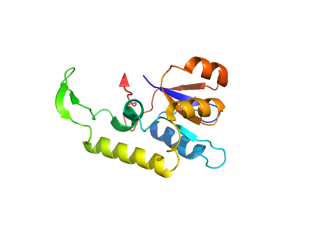
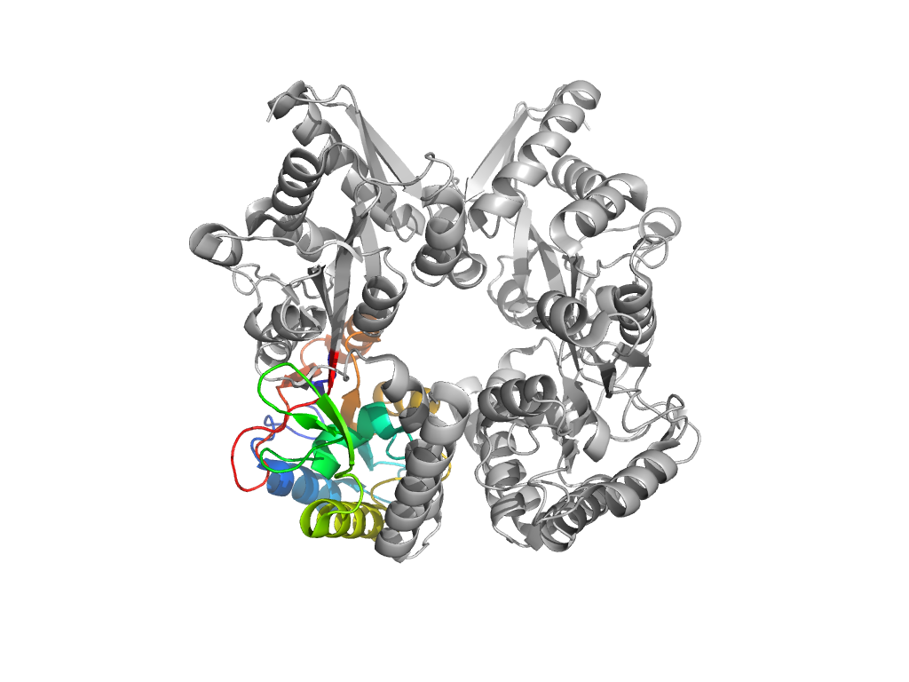
| UID: | 001520882 |
|---|---|
| Type: | Automatic domain |
| Parent: | e1ursA2 |
| Range: | A:153-303 |
| Ligands: | P33 |
| PDB: | 4hs7 |
| PDB Description: | Bacterial extracellular solute-binding protein, putative |
| UniProt: | Q2G1E9 |
| Species: |

