- A: a/b three-layered sandwiches
- X: Periplasmic binding protein-like II
- H: Periplasmic binding protein-like II
- T: Periplasmic binding protein-like II
- F: Unmapped domains
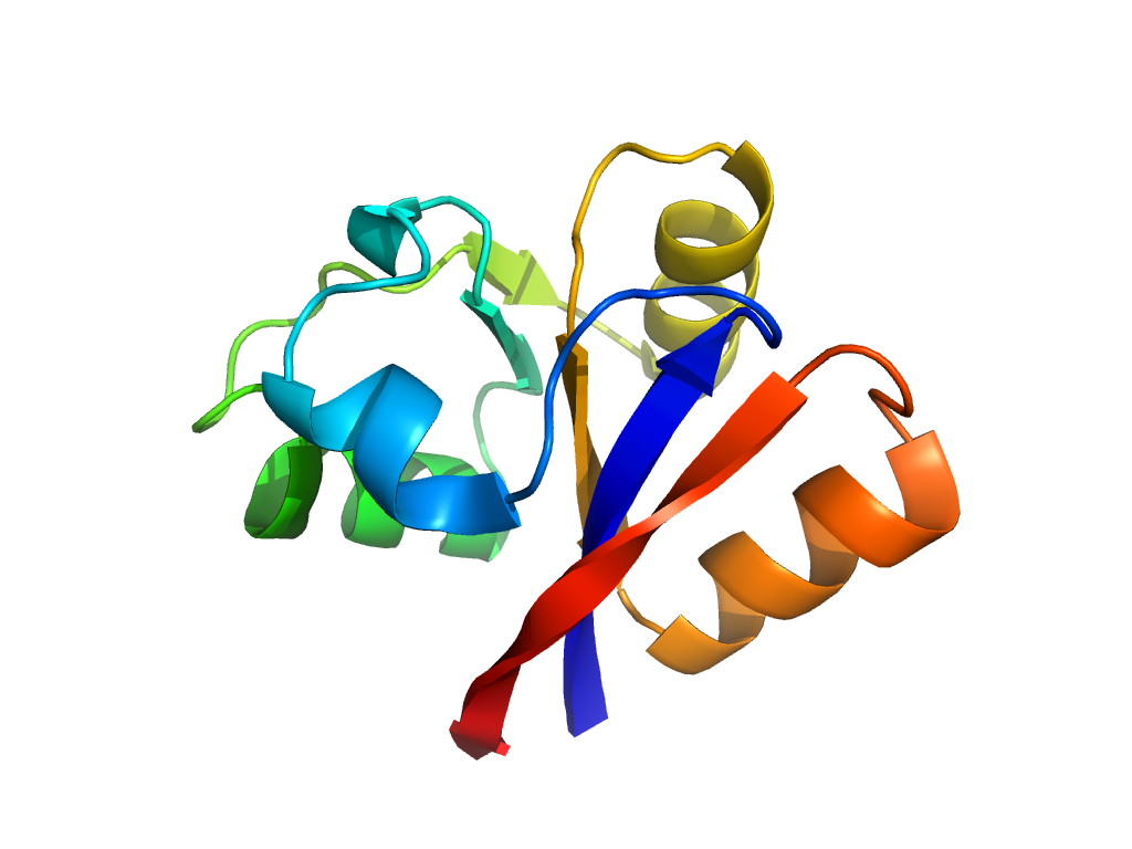
| UID: | 001521095 |
|---|---|
| Type: | Automatic domain |
| Parent: | e1ii5A3 |
| Range: | B:105-205 |
| Ligands: | ASP |
| PDB: | 4io3 |
| PDB Description: | AvGluR1 ligand binding domain |
| UniProt: | E9P5T5 |
| Species: | Adineta vaga |


