- A: a/b three-layered sandwiches
- X: Periplasmic binding protein-like II
- H: Periplasmic binding protein-like II
- T: Periplasmic binding protein-like II
- F: SBP_bac_1
| UID: | 001823212 |
|---|---|
| Type: | Automatic domain |
| Parent: | e4jbzA1 |
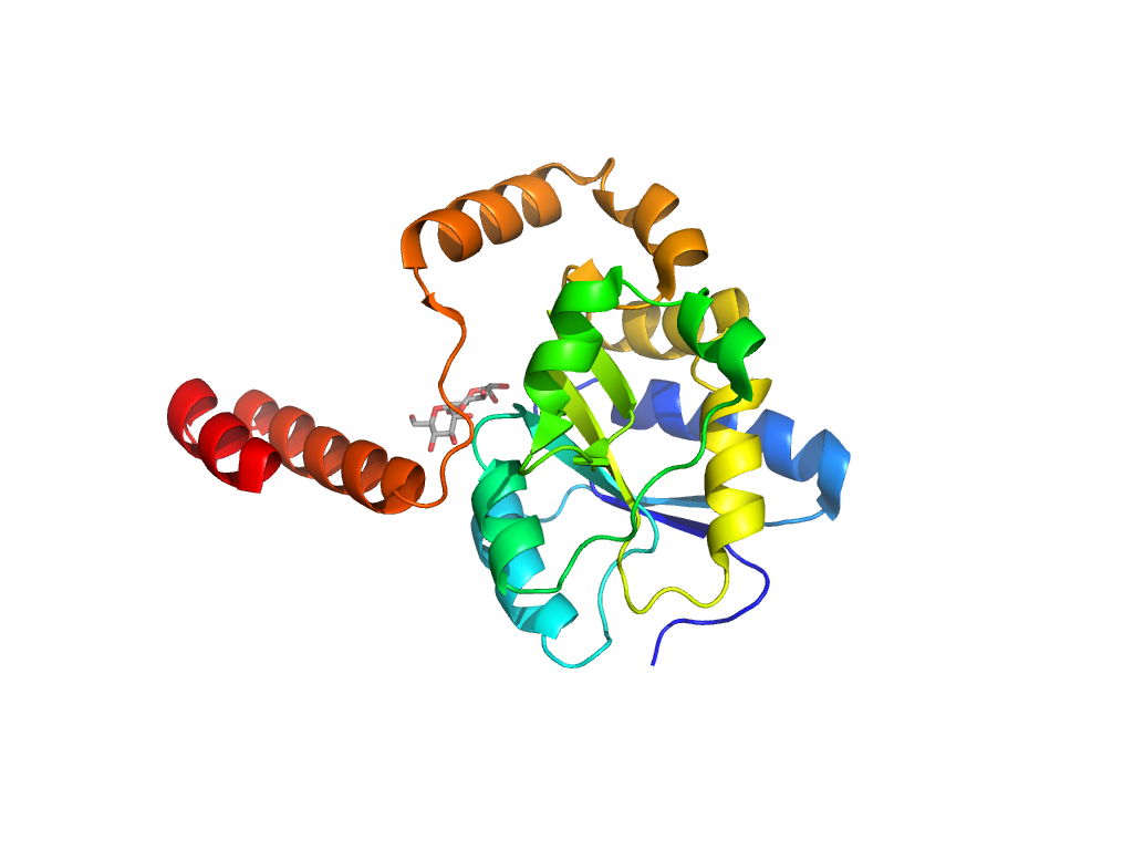
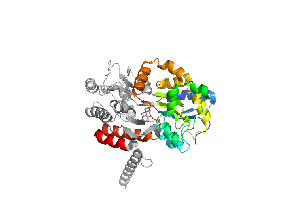
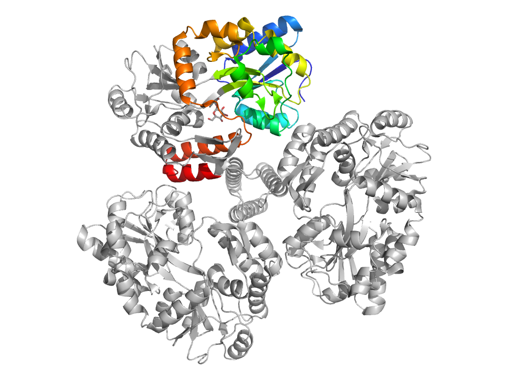
| Range: | B:1-110,B:261-366 |
| Ligands: | MAL |
| PDB: | 4jbz |
| PDB Description: | MALTOSE-BINDING PERIPLASMIC PROTEIN FUSED WITH XENOPUS LAEVIS MCM10 COILED-COIL REGION |
| UniProt: | P0AEX9 |
| Species: | Xenopus laevis |
