- A: a/b three-layered sandwiches
- X: Periplasmic binding protein-like II
- H: Periplasmic binding protein-like II
- T: Periplasmic binding protein-like II
- F: DctP
| UID: | 001523496 |
|---|---|
| Type: | Automatic domain |
| Parent: | e3gyyA2 |
| Range: | A:146-234 |
| Ligands: | 2UF SO4 |
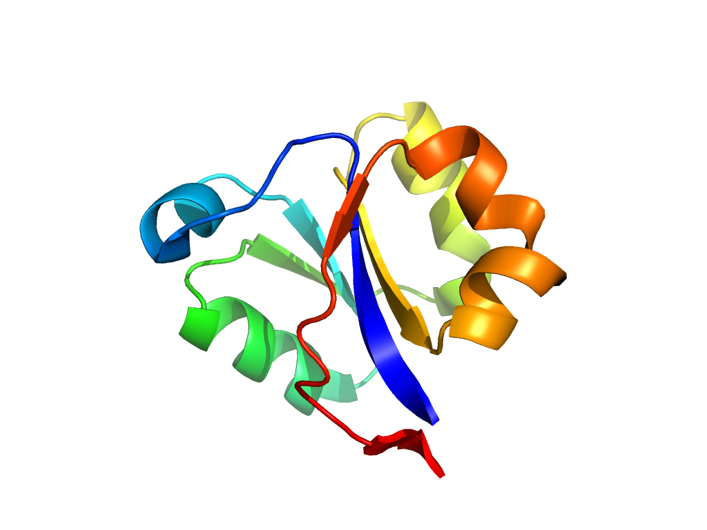
| PDB: | 4pbq |
| PDB Description: | Putative TRAP PERIPLASMIC SOLUTE BINDING PROTEIN |
| UniProt: | C9MHP2 |
| Species: |


