Domain ID e4u1gA1

Download files:
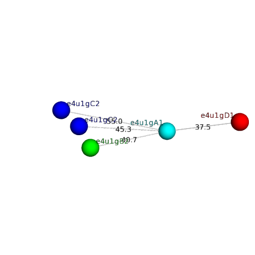
  • A: alpha duplicates or obligate multimers
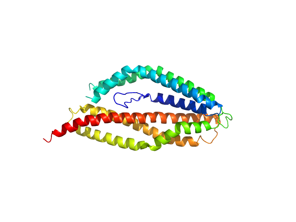
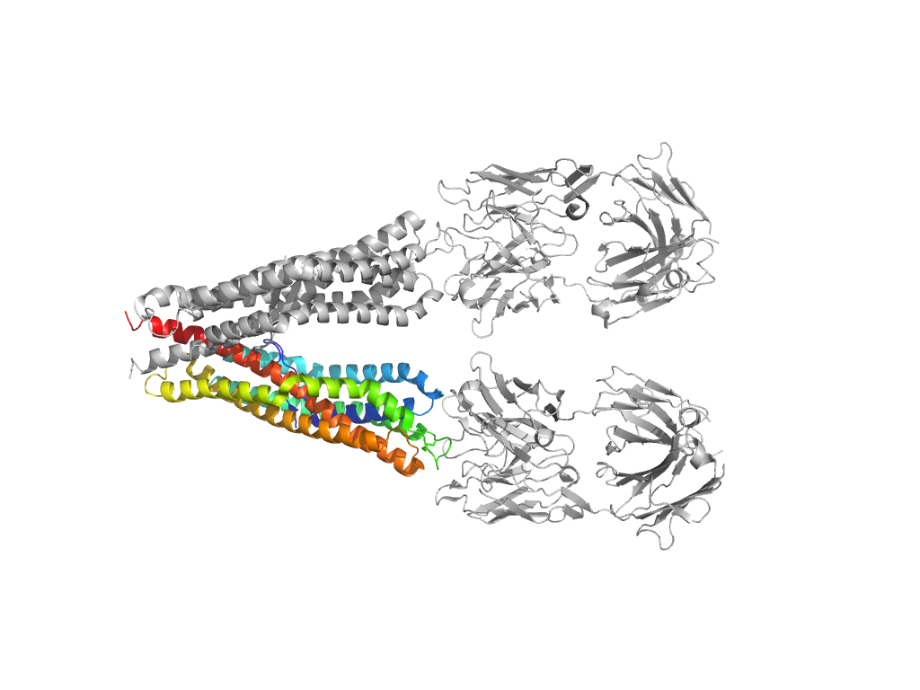
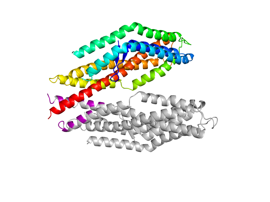
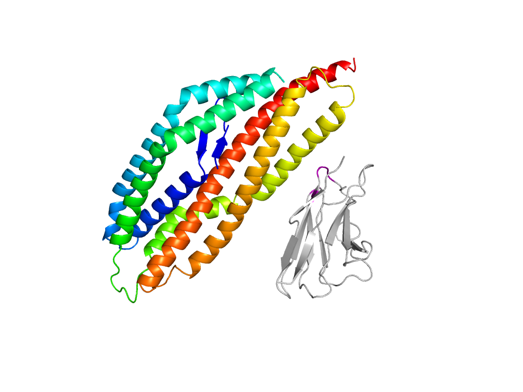
  • X: Reticulocyte binding protein 5
  • H: Reticulocyte binding protein 5
  • T: Reticulocyte binding protein 5
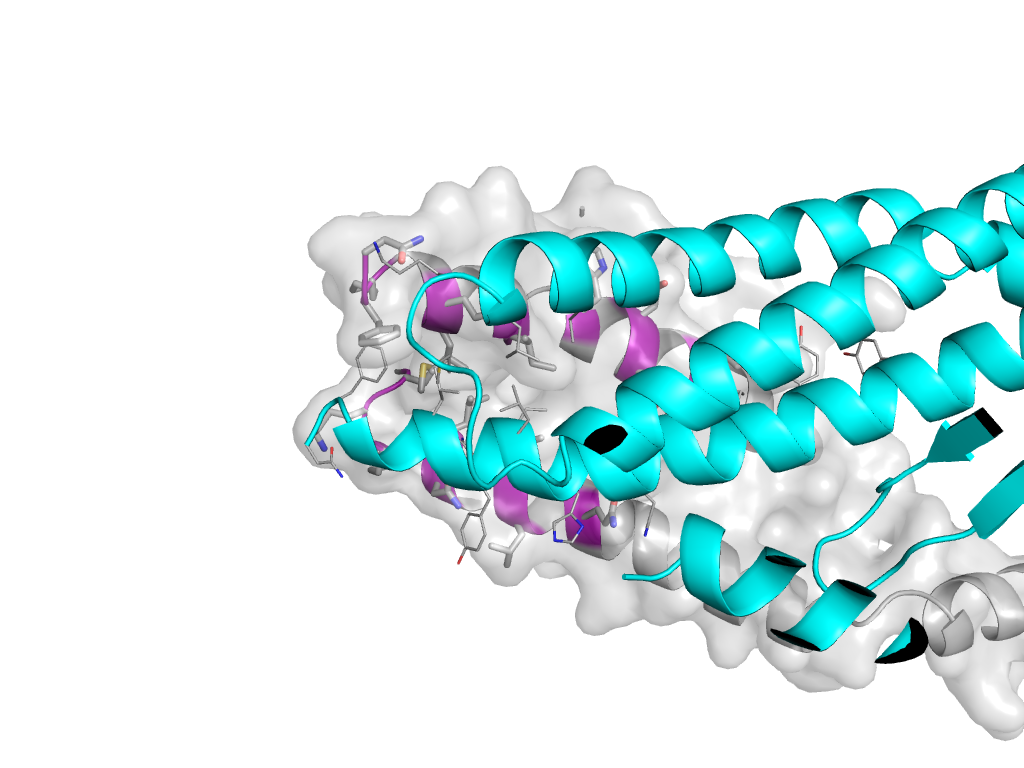
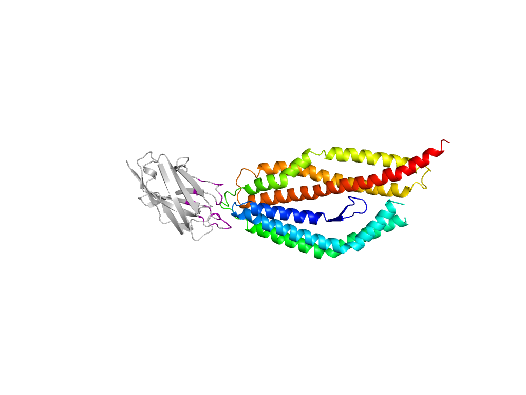
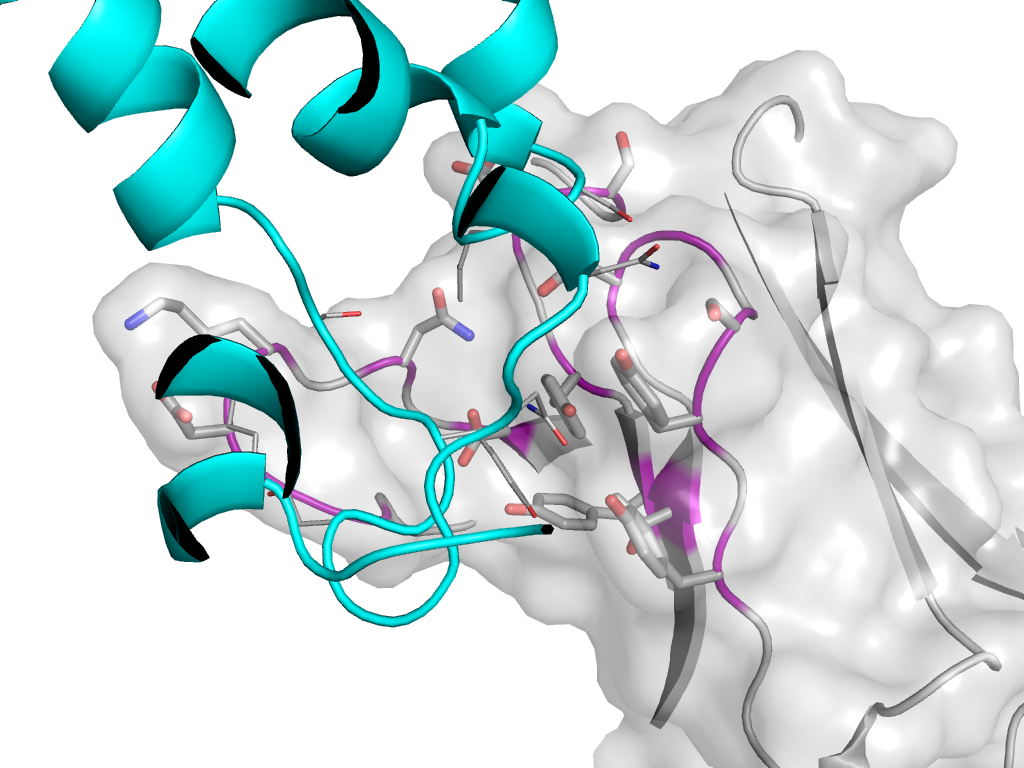
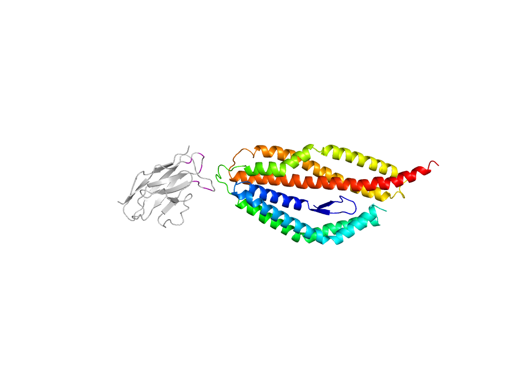
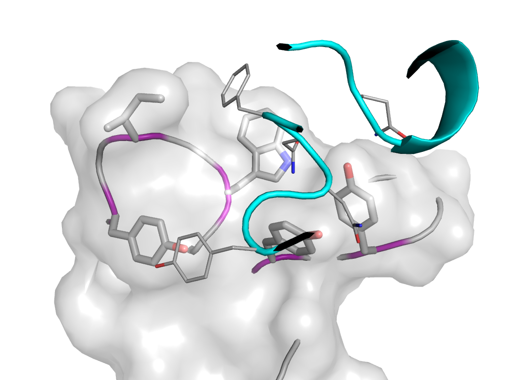
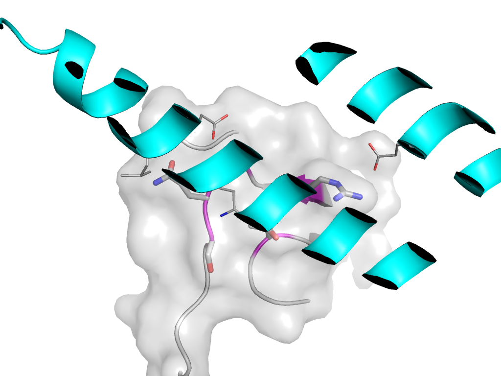
  • F: Rh5
UID: 001325238
Type: Automatic domain
Parent: e4u1gD1
Range: A:160-241,A:301-506
PDB: 4u1g
PDB Description: Reticulocyte binding protein 5
UniProt: B2L3N7
Species: Plasmodium falciparum

Structure of domain e4u1gA1

Domains in the same chain:

e4u1gA1 A:160-241,A:301-506 Reticulocyte binding protein 5 Reticulocyte binding protein 5 Reticulocyte binding protein 5

Domains in the same PDB:

e4u1gA1 A:160-241,A:301-506 Reticulocyte binding protein 5 Reticulocyte binding protein 5 Reticulocyte binding protein 5
e4u1gB2 B:3-122 Immunoglobulin-like beta-sandwich Immunoglobulin-related Immunoglobulin/Fibronectin type III/E set domains/PapD-like
e4u1gB1 B:123-221 Immunoglobulin-like beta-sandwich Immunoglobulin-related Immunoglobulin/Fibronectin type III/E set domains/PapD-like
e4u1gC2 C:1-111 Immunoglobulin-like beta-sandwich Immunoglobulin-related Immunoglobulin/Fibronectin type III/E set domains/PapD-like
e4u1gC1 C:112-215 Immunoglobulin-like beta-sandwich Immunoglobulin-related Immunoglobulin/Fibronectin type III/E set domains/PapD-like
e4u1gD1 D:160-242,D:301-506 Reticulocyte binding protein 5 Reticulocyte binding protein 5 Reticulocyte binding protein 5
e4u1gE2 E:3-122 Immunoglobulin-like beta-sandwich Immunoglobulin-related Immunoglobulin/Fibronectin type III/E set domains/PapD-like
e4u1gE1 E:123-221 Immunoglobulin-like beta-sandwich Immunoglobulin-related Immunoglobulin/Fibronectin type III/E set domains/PapD-like
e4u1gF2 F:1-111 Immunoglobulin-like beta-sandwich Immunoglobulin-related Immunoglobulin/Fibronectin type III/E set domains/PapD-like
e4u1gF1 F:112-215 Immunoglobulin-like beta-sandwich Immunoglobulin-related Immunoglobulin/Fibronectin type III/E set domains/PapD-like

Structural contacts of domain e4u1gA1 of H-group "Reticulocyte binding protein 5":

The domain and its neighboring domains (both within the asymmetric unit and to crystallographic symmetry mates) are represented by spheres and linked by lines. Distances between the center of the domain and interfaces are shown for each contact.

Domain IDSymmetry operatorH-groupVisualization
e4u1gD1 A:X,Y,Z->D:x,y,z Reticulocyte binding protein 5 Interaction Interface Pymol
e4u1gB2 A:X,Y,Z->B:x,y,z Immunoglobulin-related Interaction Interface Pymol
e4u1gC2 A:X,Y,Z->C:x,y,z Immunoglobulin-related Interaction Interface Pymol
e4u1gC2 A:-X+2,Y-1/2,-Z-1/2->C:x,y,z Immunoglobulin-related Interaction Interface Pymol
e4u1gC2 A:-X+3,Y-1/2,-Z-1/2->C:x,y,z Immunoglobulin-related Interaction Interface Pymol