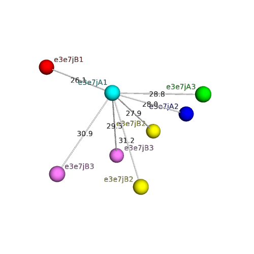
| Domain ID | Symmetry operator | H-group | Visualization |
|---|
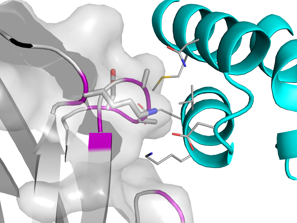
| e3e7jB1 |
A:X,Y,Z->B:x,y,z |
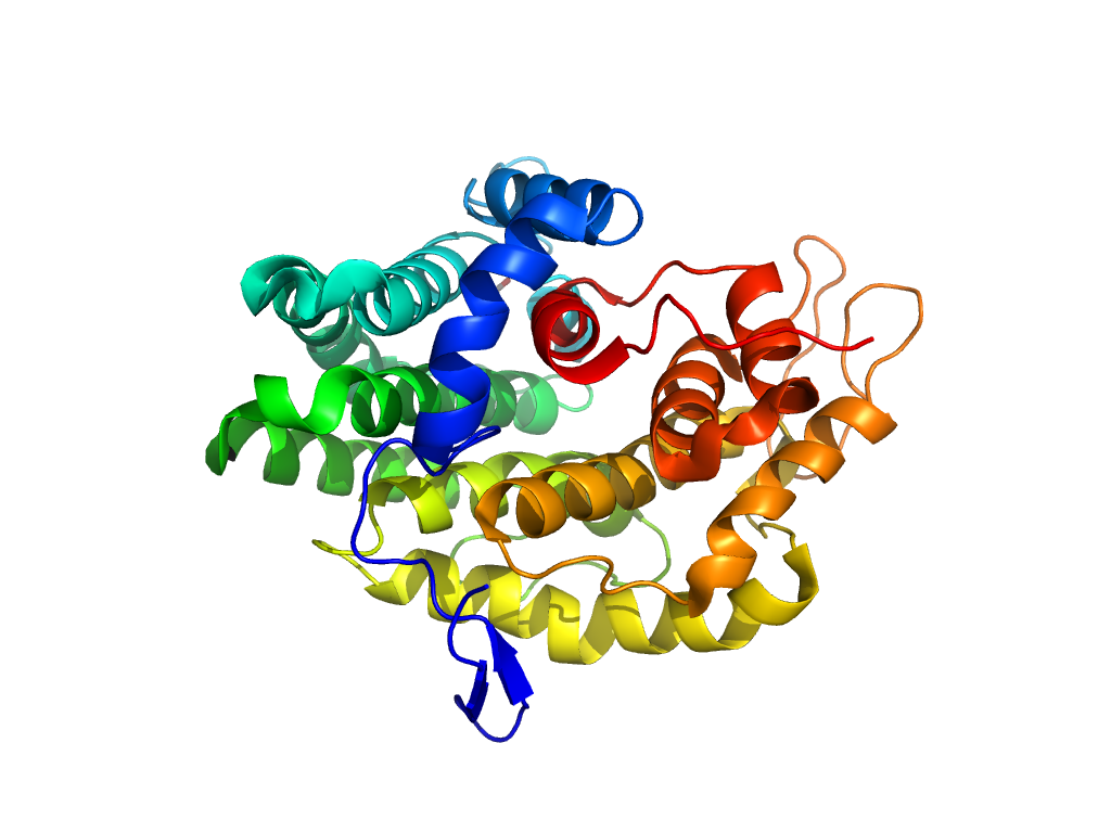
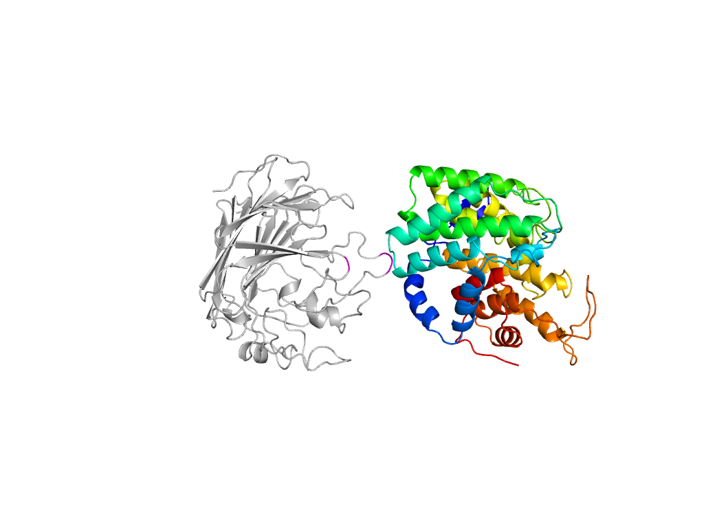
Glycosyl hydrolase domain |
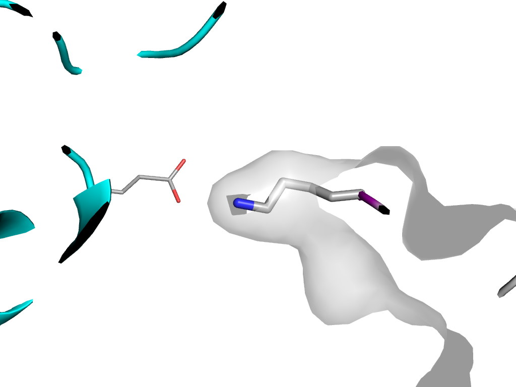
Interaction
Interface
Pymol
|
| e3e7jA3 |
A:x,y,z->A:-X+1,Y-1/2,-Z+1 |
Glycosyl hydrolase domain |
Interaction
Interface
Pymol
|
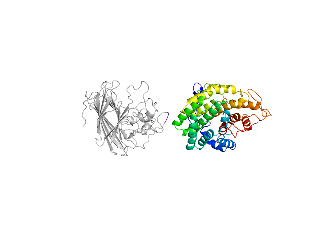
| e3e7jA2 |
A:x,y,z->A:-X+1,Y-1/2,-Z+1 |
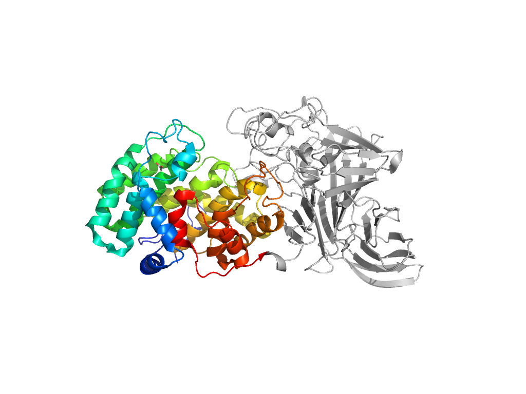
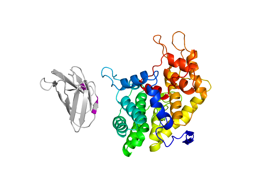
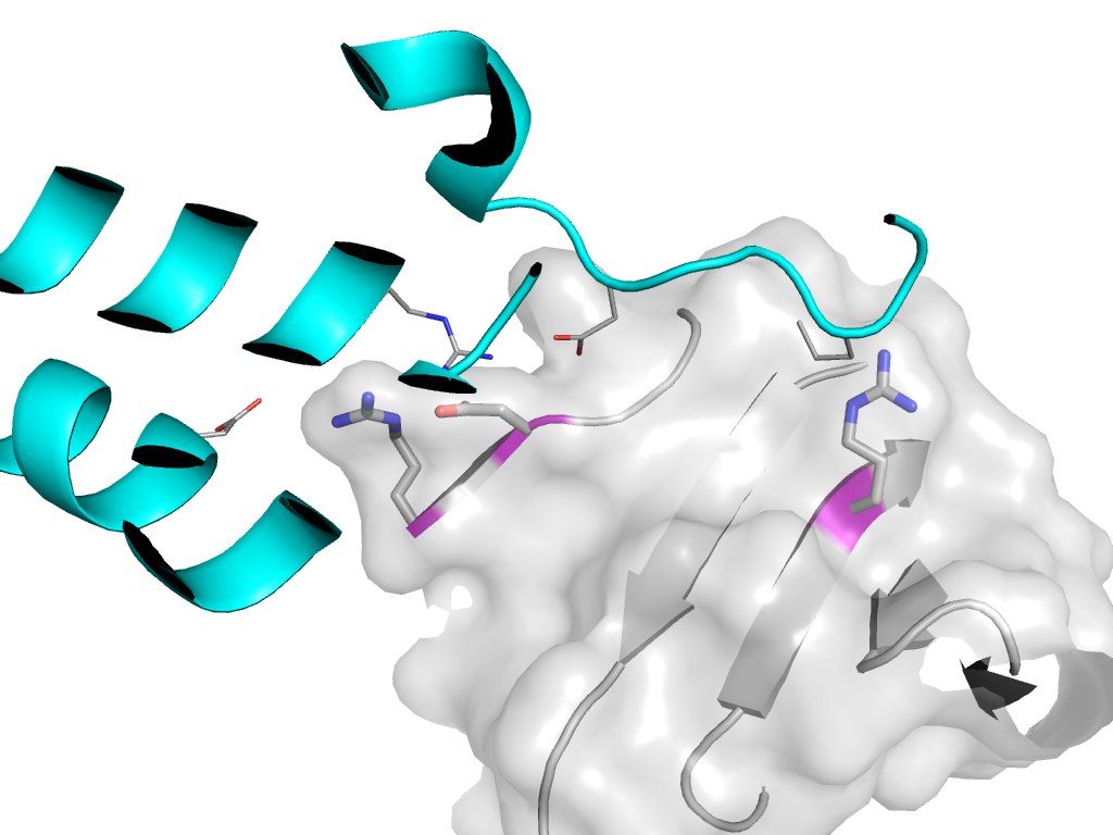
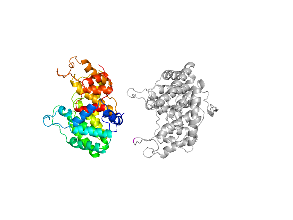
supersandwich |
Interaction
Interface
Pymol
|
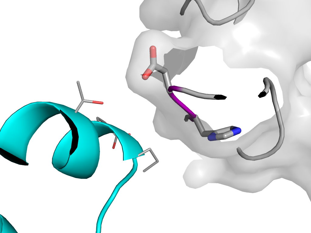
| e3e7jB2 |
A:x,y,z->B:-X,Y-1/2,-Z |
supersandwich |
Interaction
Interface
Pymol
|
| e3e7jB3 |
A:x,y,z->B:-X,Y-1/2,-Z |
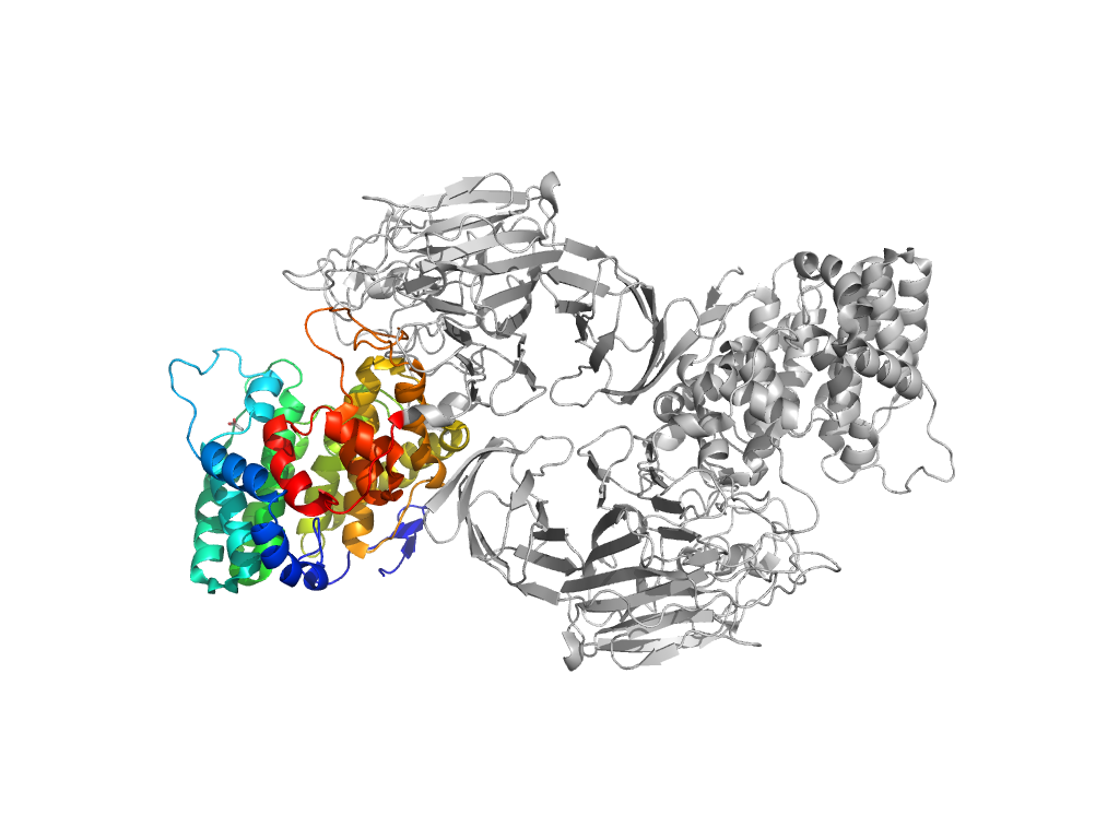
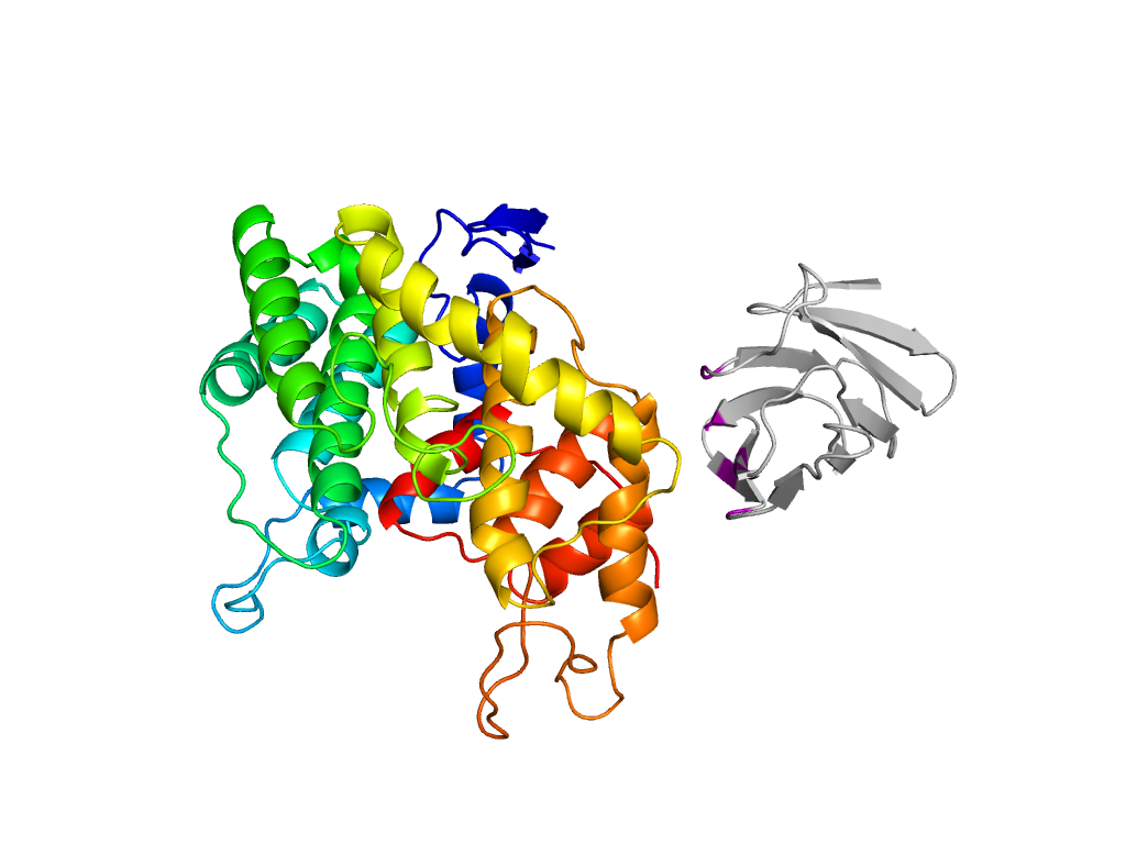
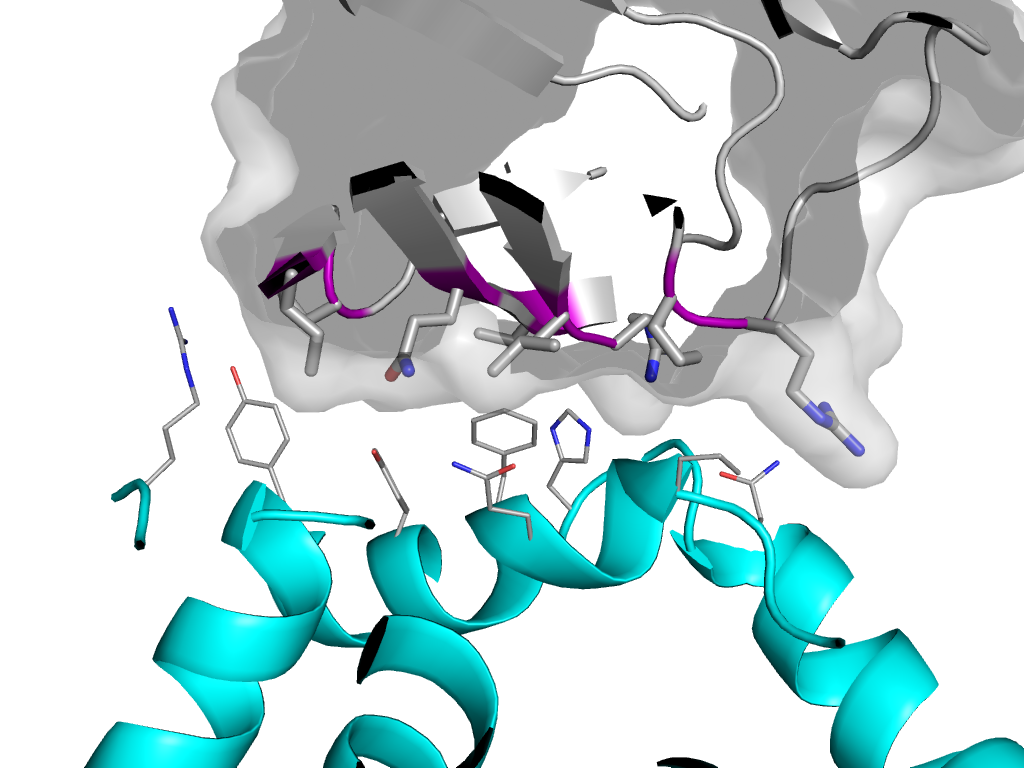
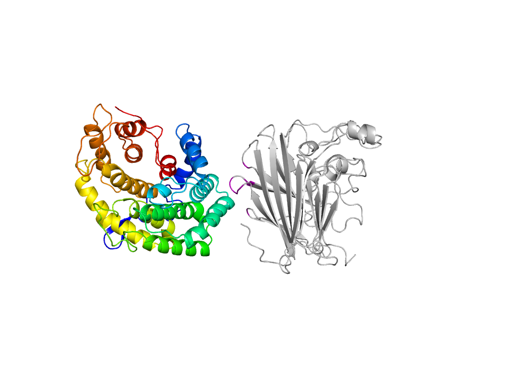
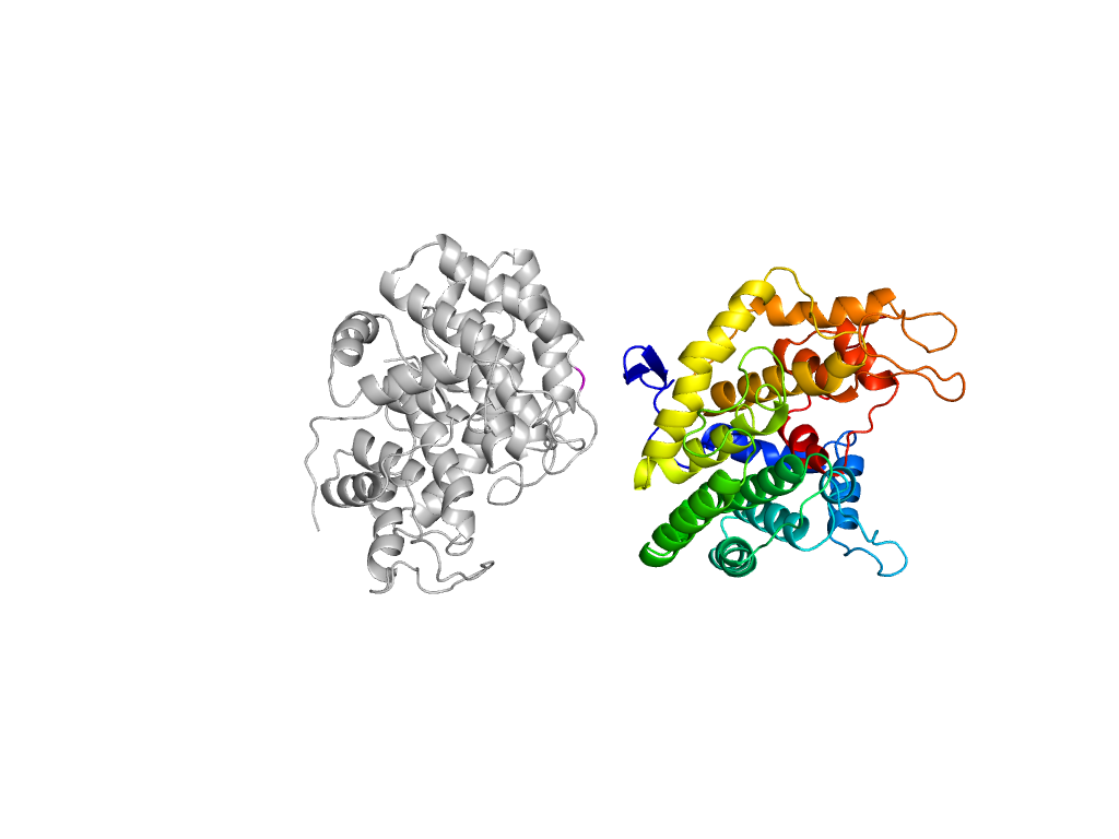
alpha/alpha toroid |
Interaction
Interface
Pymol
|
| e3e7jB3 |
A:x,y,z->B:-X+1,Y-1/2,-Z+1 |
alpha/alpha toroid |
Interaction
Interface
Pymol
|
| e3e7jB3 |
A:x,y,z->B:-X+1,Y-1/2,-Z |
alpha/alpha toroid |
Interaction
Interface
Pymol
|
| e3e7jB2 |
A:x,y,z->B:-X+1,Y-1/2,-Z |
supersandwich |
Interaction
Interface
Pymol
|