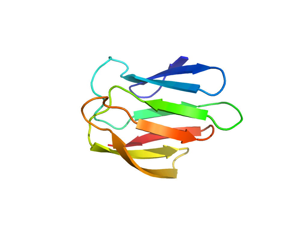
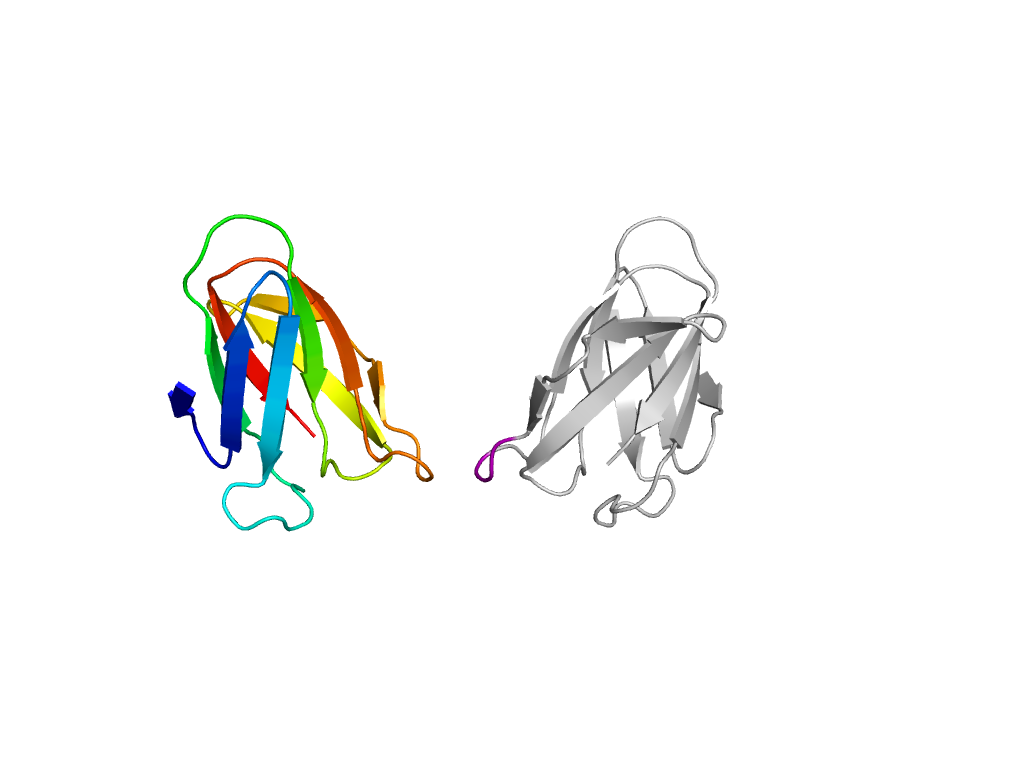
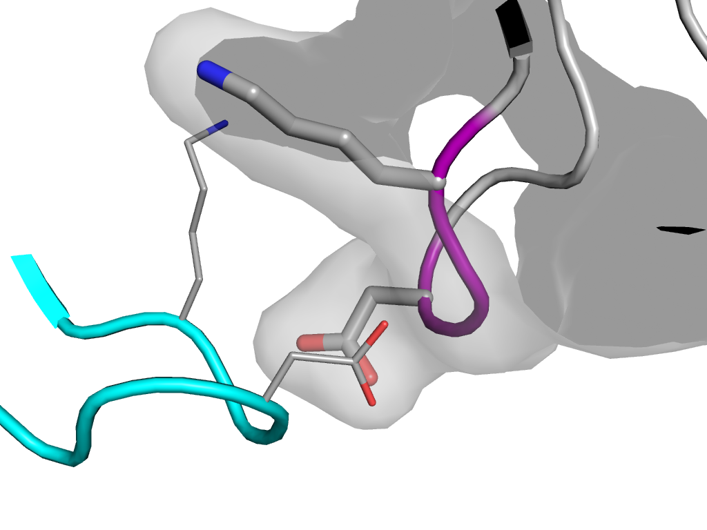
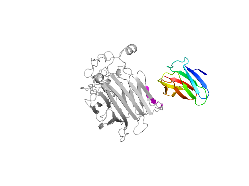
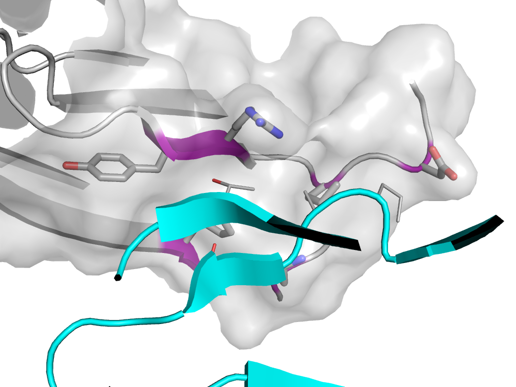
- A: beta sandwiches
- X: Glycosyl hydrolase domain-like
- H: Glycosyl hydrolase domain
- T: Glycosyl hydrolase domain
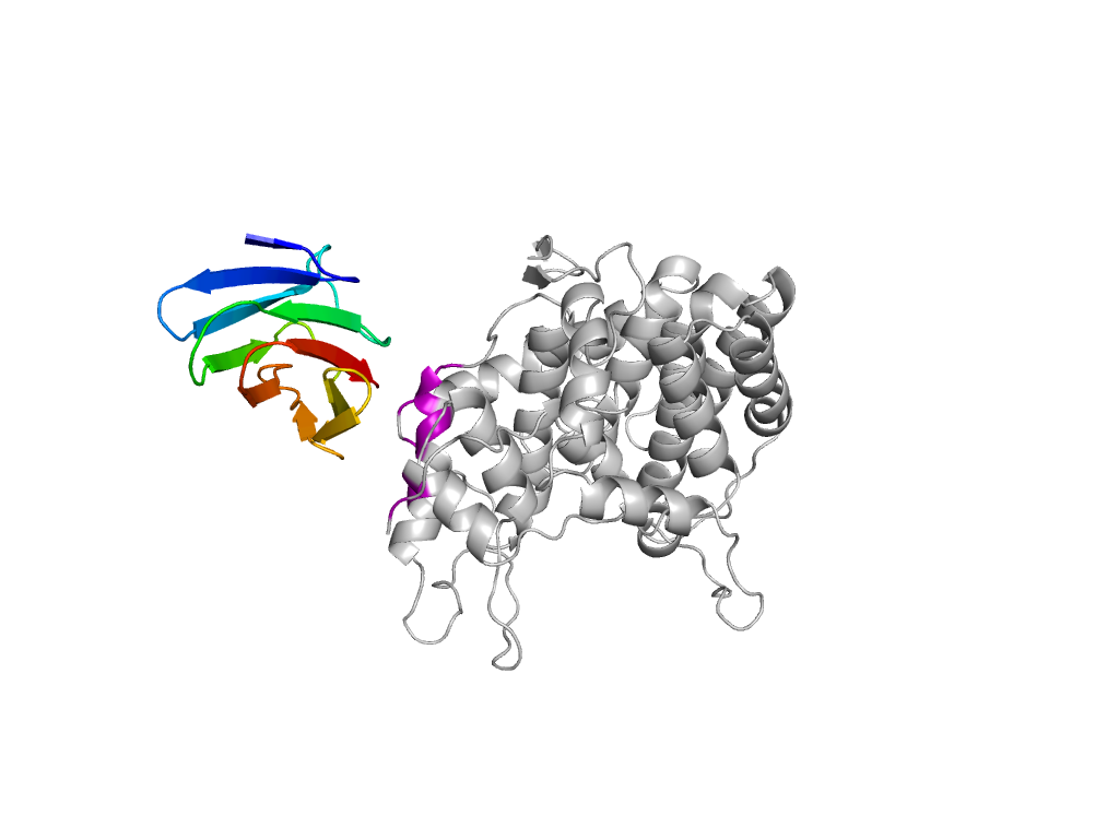
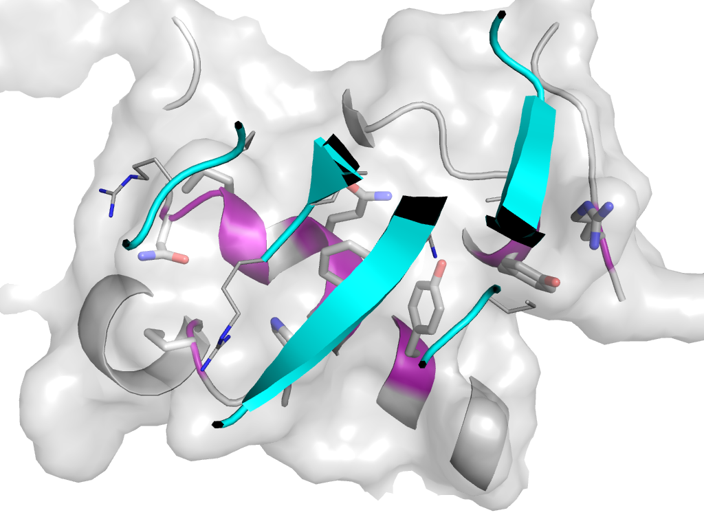
- F: HepII_C
| UID: | 000200692 |
|---|---|
| Type: | Automatic and representative domain |
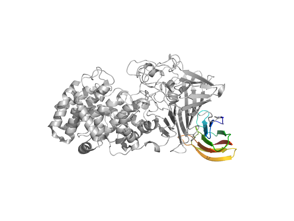
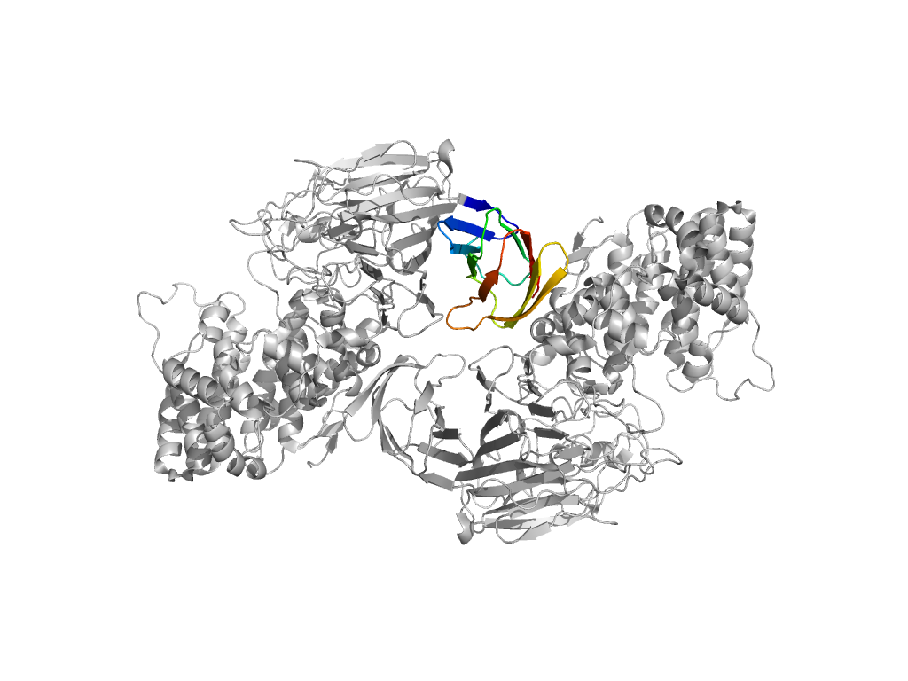
| Range: | A:686-772 |
| PDB: | 3e7j |
| PDB Description: | Heparinase II protein |
| UniProt: | C6XZB6 |
| Species: | Pedobacter heparinus |