Domain ID e3izxE1

Download files:
  • A: alpha arrays
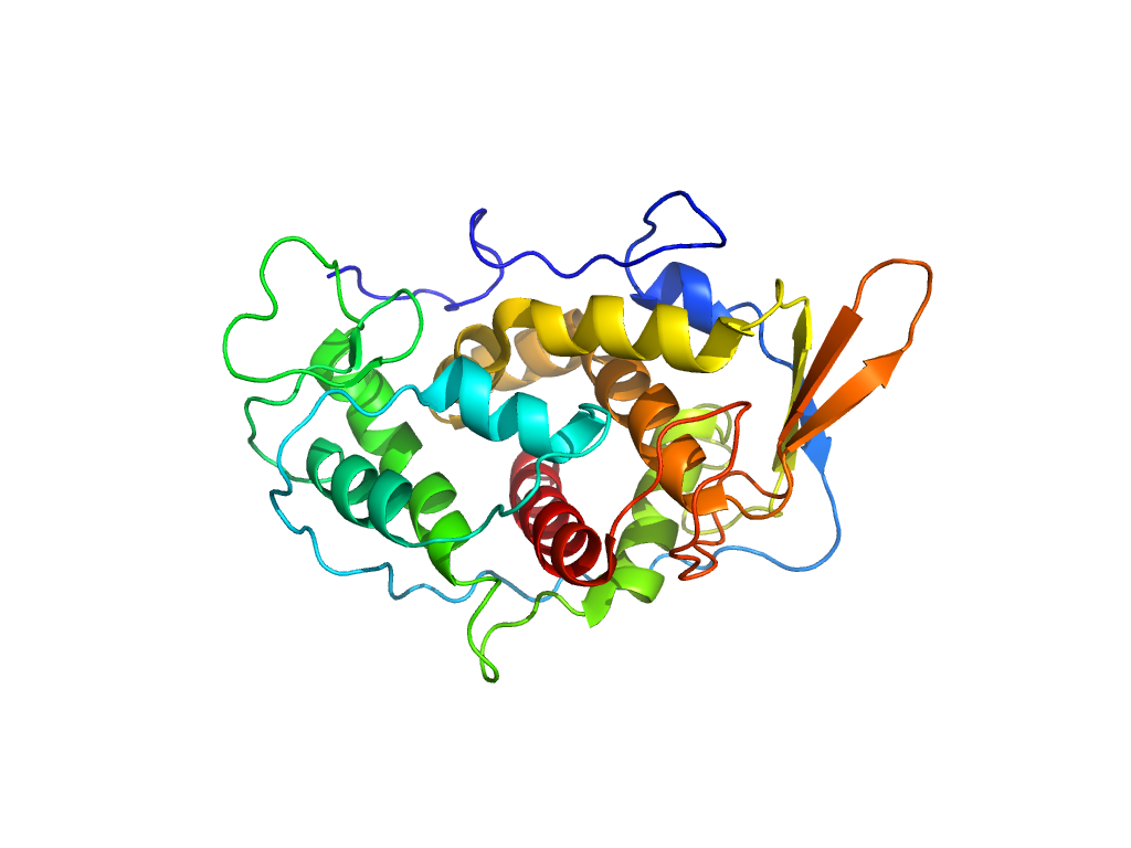
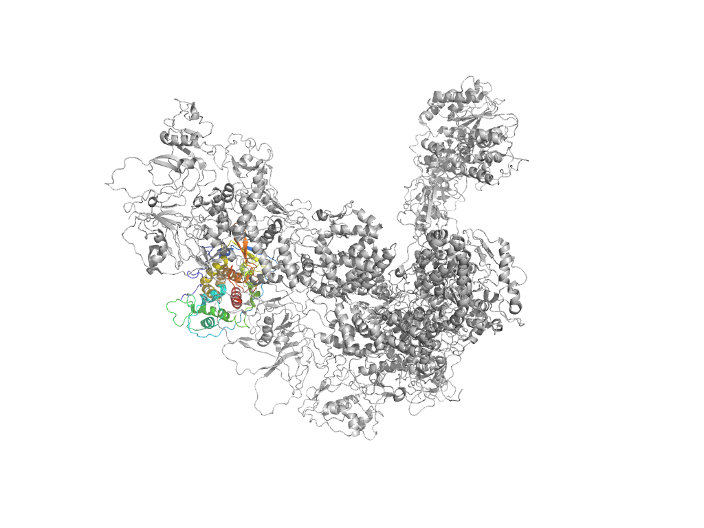
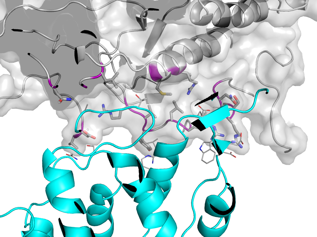
  • X: Viral structural protein 5
  • H: Viral structural protein 5
  • T: Viral structural protein 5
  • F: VP5
UID: 000422567
Type: Automatic domain
Parent: e3izxD1
Range: E:3-292
PDB: 3izx
PDB Description: Viral structural protein 5
UniProt: C6K2M8
Species:

Structure of domain e3izxE1

Domains in the same chain:

e3izxE1 E:3-292 Viral structural protein 5 Viral structural protein 5 Viral structural protein 5

Domains in the same PDB:

e3izxA3 A:1-363 Reovirus guanylyltransferase (GTase) Reovirus guanylyltransferase (GTase) Reovirus guanylyltransferase (GTase)
e3izxA4 A:364-400,A:722-821 Reovirus turret protein bridge domain Reovirus turret protein bridge domain Reovirus turret protein bridge domain
e3izxA1 A:401-721 Rossmann-like Rossmann-related S-adenosyl-L-methionine-dependent methyltransferases
e3izxA2 A:822-1057 Rossmann-like Rossmann-related S-adenosyl-L-methionine-dependent methyltransferases
e3izxB1 B:135-827,B:960-1333 Inner layer core protein p3-related Inner layer core protein p3-related Inner layer core protein p3-related
e3izxB2 B:828-959 Small protrusion domain in cytoplasmic polyhedrosis virus capsid shell protein Small protrusion domain in cytoplasmic polyhedrosis virus capsid shell protein Small protrusion domain in cytoplasmic polyhedrosis virus capsid shell protein
e3izxC1 C:74-827,C:960-1333 Inner layer core protein p3-related Inner layer core protein p3-related Inner layer core protein p3-related
e3izxC2 C:828-959 Small protrusion domain in cytoplasmic polyhedrosis virus capsid shell protein Small protrusion domain in cytoplasmic polyhedrosis virus capsid shell protein Small protrusion domain in cytoplasmic polyhedrosis virus capsid shell protein
e3izxD1 D:3-292 Viral structural protein 5 Viral structural protein 5 Viral structural protein 5
e3izxE1 E:3-292 Viral structural protein 5 Viral structural protein 5 Viral structural protein 5

Structural contacts of domain e3izxE1 of H-group "Viral structural protein 5":

The domain and its neighboring domains (both within the asymmetric unit and to crystallographic symmetry mates) are represented by spheres and linked by lines. Distances between the center of the domain and interfaces are shown for each contact.

Domain IDSymmetry operatorH-groupVisualization
e3izxC1 E:X,Y,Z->C:x,y,z Inner layer core protein p3-related Interaction Interface Pymol
e3izxB1 E:X,Y,Z->B:x,y,z Inner layer core protein p3-related Interaction Interface Pymol