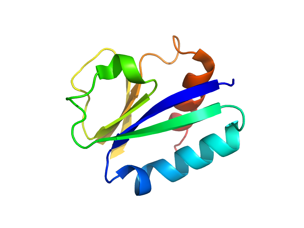
- A: a/b three-layered sandwiches
- X: Periplasmic binding protein-like II
- H: Periplasmic binding protein-like II
- T: Periplasmic binding protein-like II
- F: LysR_substrate
| UID: | 001806519 |
|---|---|
| Type: | Manual and representative domain |
| Range: | A:89-159,A:263-290 |
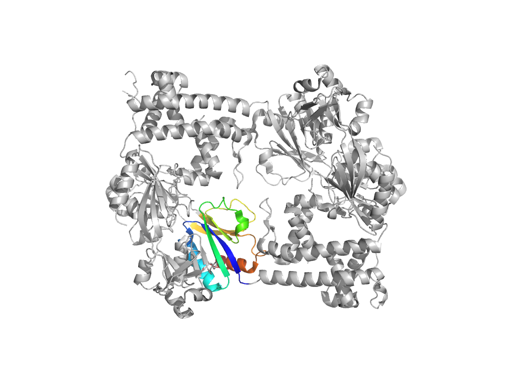
| PDB: | 3t1b |
| PDB Description: | Transcriptional regulator, LysR family |
| UniProt: | Q9KT56 |
| Species: | Vibrio cholerae |

