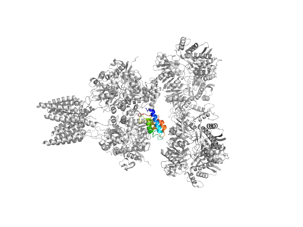
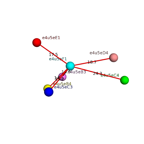
| e4u5eA1 |
A:6-111,A:244-382 |
Flavodoxin-like |
Class I glutamine amidotransferase-like |
Periplasmic binding protein-like I |
| e4u5eA2 |
A:112-243 |
Flavodoxin-like |
Class I glutamine amidotransferase-like |
Periplasmic binding protein-like I |
| e4u5eA4 |
A:383-497,A:731-789 |
Periplasmic binding protein-like II |
Periplasmic binding protein-like II |
Periplasmic binding protein-like II |
| e4u5eA3 |
A:498-505,A:632-730 |
Periplasmic binding protein-like II |
Periplasmic binding protein-like II |
Periplasmic binding protein-like II |
| e4u5eA5 |
A:506-542,A:597-627,A:790-814 |
Voltage-gated ion channels |
Voltage-gated ion channels |
Voltage-gated ion channels |
| e4u5eB2 |
B:6-111,B:244-383 |
Flavodoxin-like |
Class I glutamine amidotransferase-like |
Periplasmic binding protein-like I |
| e4u5eB1 |
B:112-243 |
Flavodoxin-like |
Class I glutamine amidotransferase-like |
Periplasmic binding protein-like I |
| e4u5eB4 |
B:384-497,B:731-789 |
Periplasmic binding protein-like II |
Periplasmic binding protein-like II |
Periplasmic binding protein-like II |
| e4u5eB5 |
B:498-505,B:632-730 |
Periplasmic binding protein-like II |
Periplasmic binding protein-like II |
Periplasmic binding protein-like II |
| e4u5eB3 |
B:506-542,B:597-627,B:790-814 |
Voltage-gated ion channels |
Voltage-gated ion channels |
Voltage-gated ion channels |
| e4u5eC5 |
C:6-111,C:244-381 |
Flavodoxin-like |
Class I glutamine amidotransferase-like |
Periplasmic binding protein-like I |
| e4u5eC3 |
C:112-243 |
Flavodoxin-like |
Class I glutamine amidotransferase-like |
Periplasmic binding protein-like I |
| e4u5eC4 |
C:382-497,C:731-789 |
Periplasmic binding protein-like II |
Periplasmic binding protein-like II |
Periplasmic binding protein-like II |
| e4u5eC1 |
C:498-505,C:632-730 |
Periplasmic binding protein-like II |
Periplasmic binding protein-like II |
Periplasmic binding protein-like II |
| e4u5eC2 |
C:506-542,C:597-627,C:790-814 |
Voltage-gated ion channels |
Voltage-gated ion channels |
Voltage-gated ion channels |
| e4u5eD1 |
D:6-111,D:244-381 |
Flavodoxin-like |
Class I glutamine amidotransferase-like |
Periplasmic binding protein-like I |
| e4u5eD4 |
D:112-243 |
Flavodoxin-like |
Class I glutamine amidotransferase-like |
Periplasmic binding protein-like I |
| e4u5eD3 |
D:382-497,D:731-789 |
Periplasmic binding protein-like II |
Periplasmic binding protein-like II |
Periplasmic binding protein-like II |
| e4u5eD2 |
D:498-505,D:632-730 |
Periplasmic binding protein-like II |
Periplasmic binding protein-like II |
Periplasmic binding protein-like II |
| e4u5eD5 |
D:506-542,D:597-627,D:790-814 |
Voltage-gated ion channels |
Voltage-gated ion channels |
Voltage-gated ion channels |
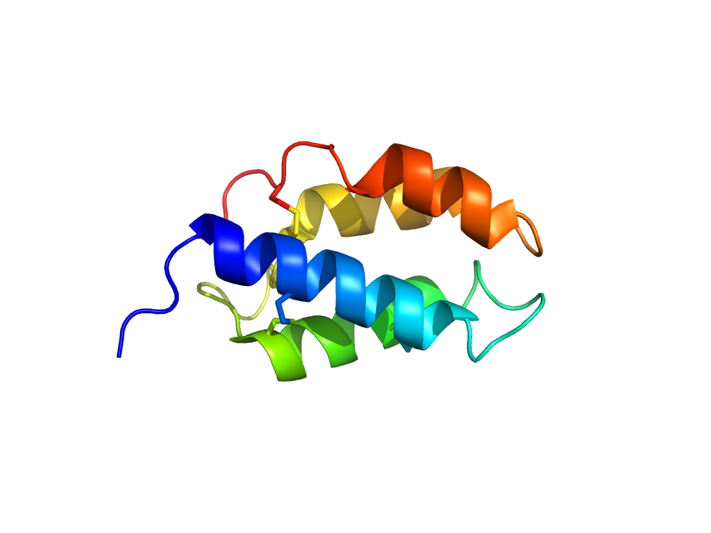
| e4u5eE1 |
E:2-86 |
con-ikot-ikot toxin |
con-ikot-ikot toxin |
con-ikot-ikot toxin |
| e4u5eF1 |
F:2-86 |
con-ikot-ikot toxin |
con-ikot-ikot toxin |
con-ikot-ikot toxin |
