Domain ID e3oxnC2

Download files:
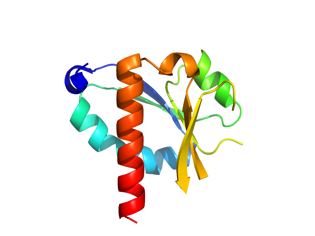
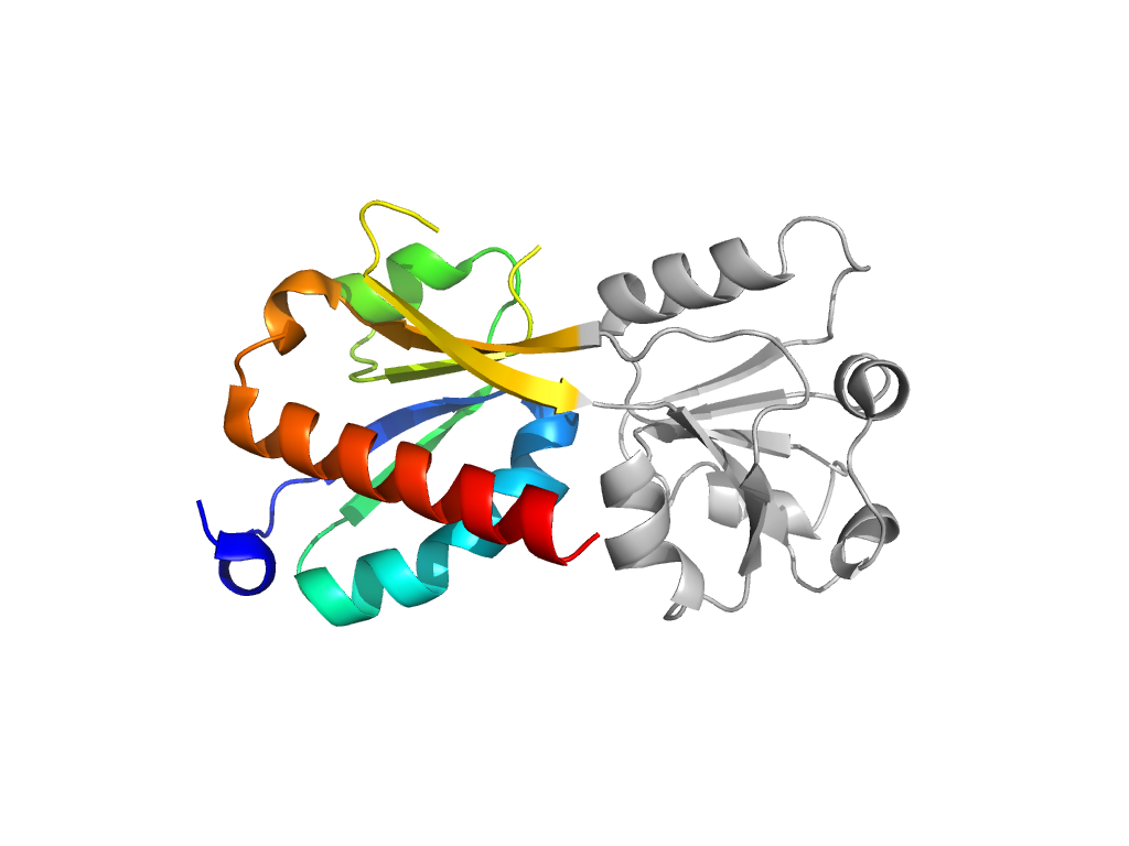
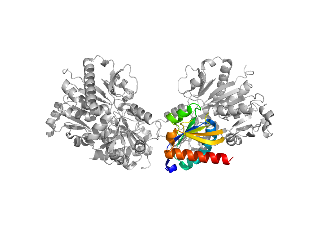
  • A: a/b three-layered sandwiches
  • X: Periplasmic binding protein-like II
  • H: Periplasmic binding protein-like II
  • T: Periplasmic binding protein-like II
  • F: LysR_substrate
UID: 001522075
Type: Automatic domain
Parent: e3t1bA2
Range: C:109-187,C:290-323
PDB: 3oxn
PDB Description: Putative transcriptional regulator, LysR family
UniProt: Q87TP2
Species: Vibrio parahaemolyticus

Structure of domain e3oxnC2

Structural contacts of domain e3oxnC2 of H-group "Periplasmic binding protein-like II":

Sorry, the images are being generated.

The domain and its neighboring domains (both within the asymmetric unit and to crystallographic symmetry mates) are represented by spheres and linked by lines. Distances between the center of the domain and interfaces are shown for each contact.

Domain IDSymmetry operatorH-groupVisualization
e3oxnB1 C:x,y,z->B:X,Y,Z Periplasmic binding protein-like II Interaction Interface Pymol
e3oxnB2 C:x,y,z->B:X,Y,Z Periplasmic binding protein-like II Interaction Interface Pymol
e3oxnB1 C:x,y,z->B:X,Y-1,Z Periplasmic binding protein-like II Interaction Interface Pymol
e3oxnA2 C:x,y,z->A:X,Y,Z Periplasmic binding protein-like II Interaction Interface Pymol
e3oxnD2 C:X,Y,Z->D:x,y,z Periplasmic binding protein-like II Interaction Interface Pymol